

Increased potency of the PHSCN dendrimer as an inhibitor of human prostate cancer cell invasion, extravasation, and lung colony formation
- PMID: 20339907
- PMCID: PMC3053599
- DOI: 10.1007/s10585-010-9316-1
Increased potency of the PHSCN dendrimer as an inhibitor of human prostate cancer cell invasion, extravasation, and lung colony formation
Abstract
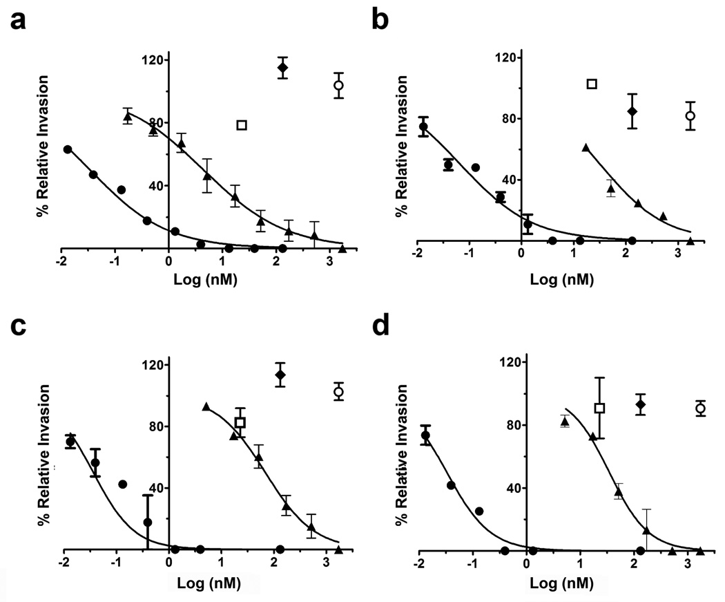
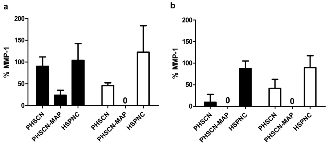
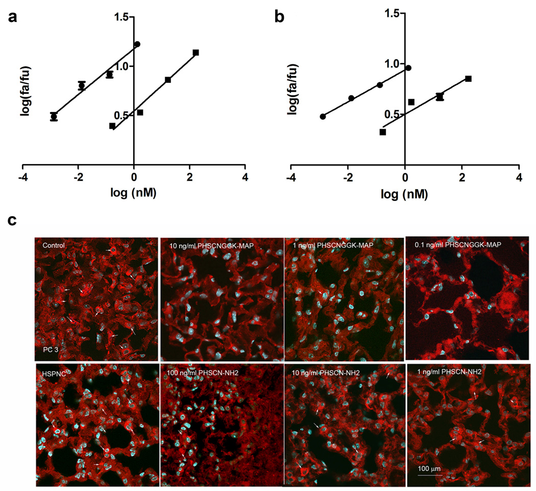
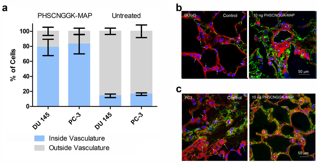
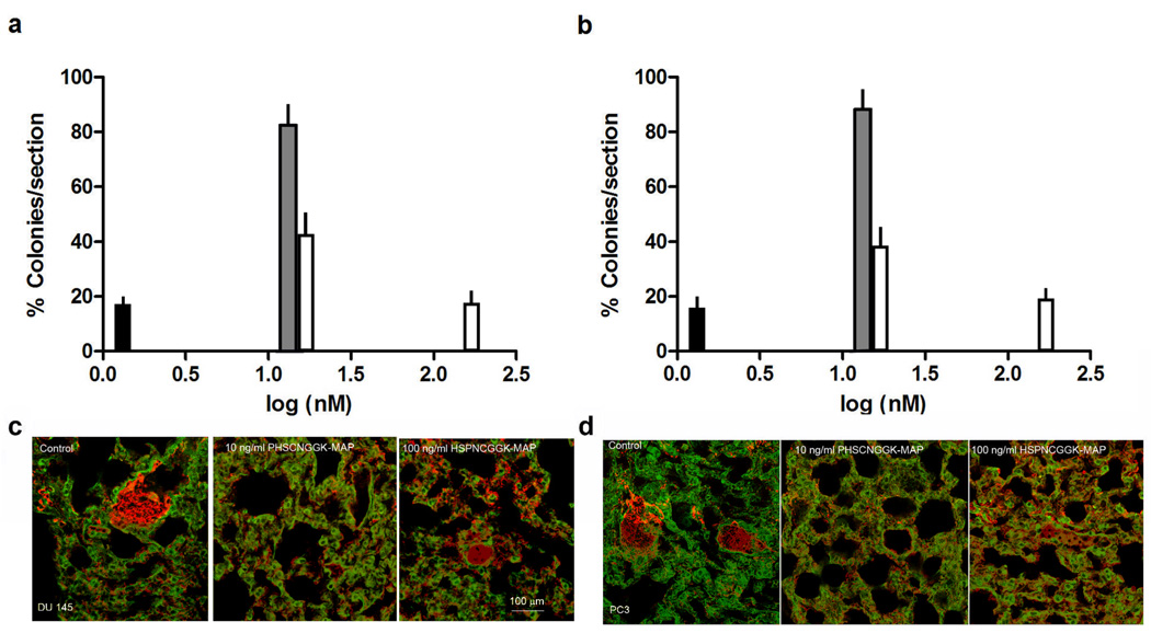
Activated alpha5beta1 integrin occurs specifically on tumor cells and on endothelial cells of tumor-associated vasculature, and plays a key role in invasion and metastasis. The PHSCN peptide (Ac-PHSCN-NH(2)) preferentially binds activated alpha5beta1, to block invasion in vitro, and inhibit growth, metastasis and tumor recurrence in preclinical models of prostate cancer. In Phase I clinical trial, systemic Ac-PHSCN-NH(2) monotherapy was well tolerated, and metastatic disease progression was prevented for 4-14 months in one-third of treated patients. We have developed a significantly more potent derivative, the PHSCN-polylysine dendrimer (Ac-PHSCNGGK-MAP). Using in vitro invasion assays with naturally serum-free basement membranes, we observed that the PHSCN dendrimer was 130- to 1900-fold more potent than the PHSCN peptide at blocking alpha5beta1-mediated invasion by DU 145 and PC-3 human prostate cancer cells, whether invasion was induced by serum, or by the Ac-PHSRN-NH(2) peptide, under serum-free conditions. The PHSCN dendrimer was also approximately 800 times more effective than PHSCN peptide at preventing DU 145 and PC-3 extravasation in the lungs of athymic mice. Chou-Talalay analysis suggested that inhibition of both invasion in vitro and extravasation in vivo by the PHSCN dendrimer are highly synergistic. We found that many extravasated DU 145 and PC-3 cells go onto develop into metastatic colonies, and that a single pretreatment with the PHSCN dendrimer was 100-fold more affective than the PHSCN peptide at reducing lung colony formation. Since many patients newly diagnosed with prostate cancer already have locally advanced or metastatic disease, the availability of a well-tolerated, nontoxic systemic therapy, like the PHSCN dendrimer, which prevents metastatic progression by inhibiting invasion, could be very beneficial.
Figures

Similar articles
-
A D-amino acid containing peptide as a potent, noncovalent inhibitor of α5β1 integrin in human prostate cancer invasion and lung colonization.Clin Exp Metastasis. 2014 Apr;31(4):379-93. doi: 10.1007/s10585-013-9634-1. Epub 2014 Jan 25. Clin Exp Metastasis. 2014. PMID: 24464034
-
The PHSCN dendrimer as a more potent inhibitor of human breast cancer cell invasion, extravasation, and lung colony formation.Breast Cancer Res Treat. 2011 Jan;125(2):363-75. doi: 10.1007/s10549-010-0826-y. Epub 2010 Mar 19. Breast Cancer Res Treat. 2011. PMID: 20300829
-
Therapeutic inhibition of breast cancer bone metastasis progression and lung colonization: breaking the vicious cycle by targeting α5β1 integrin.Breast Cancer Res Treat. 2016 Jun;157(3):489-501. doi: 10.1007/s10549-016-3844-6. Epub 2016 Jun 2. Breast Cancer Res Treat. 2016. PMID: 27255534
-
Suppression of tumor recurrence and metastasis by a combination of the PHSCN sequence and the antiangiogenic compound tetrathiomolybdate in prostate carcinoma.Neoplasia. 2002 Sep-Oct;4(5):373-9. doi: 10.1038/sj.neo.7900258. Neoplasia. 2002. PMID: 12192595 Free PMC article.
-
Targeting invasion induction as a therapeutic strategy for the treatment of cancer.Curr Cancer Drug Targets. 2005 Nov;5(7):489-503. doi: 10.2174/156800905774574002. Curr Cancer Drug Targets. 2005. PMID: 16305346 Review.
Cited by
-
A D-amino acid containing peptide as a potent, noncovalent inhibitor of α5β1 integrin in human prostate cancer invasion and lung colonization.Clin Exp Metastasis. 2014 Apr;31(4):379-93. doi: 10.1007/s10585-013-9634-1. Epub 2014 Jan 25. Clin Exp Metastasis. 2014. PMID: 24464034
-
RGD-Binding Integrins in Prostate Cancer: Expression Patterns and Therapeutic Prospects against Bone Metastasis.Cancers (Basel). 2012 Oct 26;4(4):1106-45. doi: 10.3390/cancers4041106. Cancers (Basel). 2012. PMID: 24213501 Free PMC article.
-
Targeting integrin α5β1 in urological tumors: opportunities and challenges.Front Oncol. 2023 Jul 6;13:1165073. doi: 10.3389/fonc.2023.1165073. eCollection 2023. Front Oncol. 2023. PMID: 37483505 Free PMC article. Review.
-
Bone Metastasis from Renal Cell Carcinoma.Int J Mol Sci. 2016 Jun 22;17(6):987. doi: 10.3390/ijms17060987. Int J Mol Sci. 2016. PMID: 27338367 Free PMC article. Review.
-
Integrin α5β1, the Fibronectin Receptor, as a Pertinent Therapeutic Target in Solid Tumors.Cancers (Basel). 2013 Jan 15;5(1):27-47. doi: 10.3390/cancers5010027. Cancers (Basel). 2013. PMID: 24216697 Free PMC article.
References
-
- Livant DL, Brabec RK, Pienta KJ, Allen DL, Kurachi K, Markwart S, Upadhyaya A. Anti-invasive, antitumorigenic, and antimetastatic activities of the PHSCN sequence in prostate carcinoma. Cancer Res. 2000;60:309–320. - PubMed
-
- Zeng ZZ, Jia Y, Hahn NJ, Markwart SM, Rockwood KF, Livant DL. Role of focal adhesion kinase and phosphatidylinositol 3'-kinase in integrin fibronectin receptor-mediated, matrix metalloproteinase-1-dependent invasion by metastatic prostate cancer cells. Cancer Res. 2006;66:8091–8099. - PubMed
-
- Fornaro M, Manes T, Languino LR. Integrins and prostate cancer metastases. Cancer Metastasis Rev. 2001;20:321–331. - PubMed
-
- Miles FL, Pruitt FL, van Golen KL, Cooper CR. Stepping out of the flow: capillary extravasation in cancer metastasis. Clin Exp Metastasis. 2008;25:305–324. - PubMed
Publication types
MeSH terms
Substances
Grants and funding
LinkOut - more resources
Full Text Sources
Other Literature Sources
Medical
