. 2010 Nov;64(11):829-38.
doi: 10.1002/syn.20793.

ER-β mediates 17β-estradiol attenuation of HIV-1 Tat-induced apoptotic signaling

Affiliations

ER-β mediates 17β-estradiol attenuation of HIV-1 Tat-induced apoptotic signaling

Sheila M Adams et al. Synapse. 2010 Nov.

Abstract

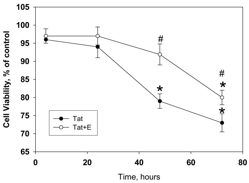
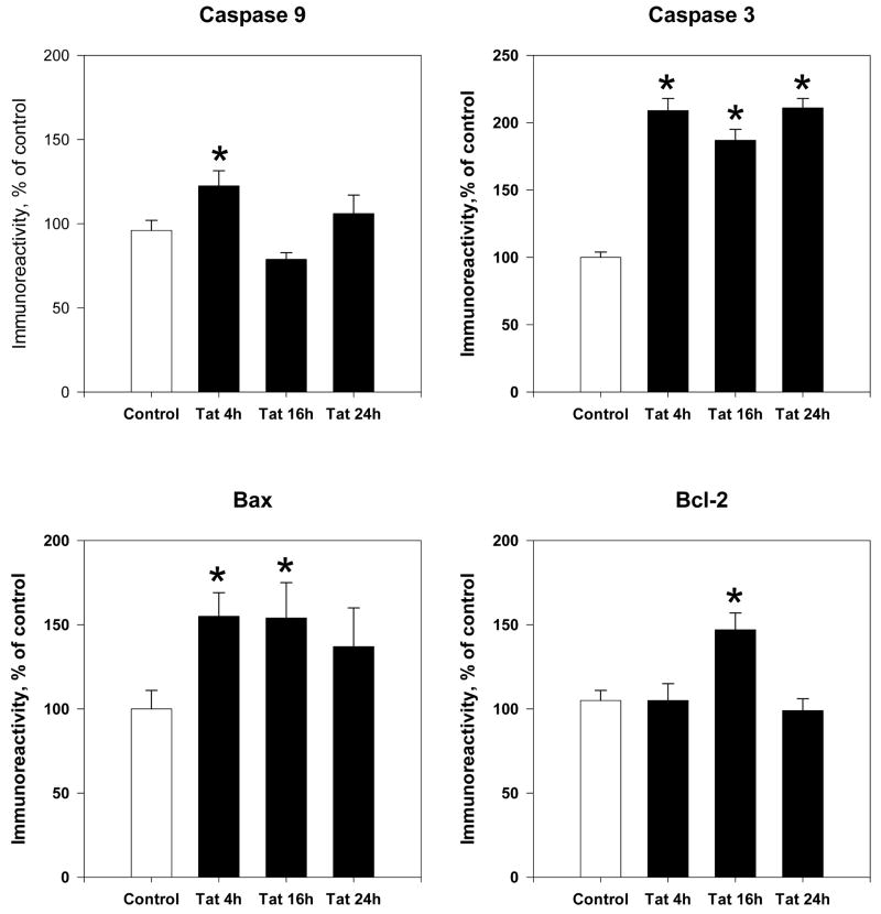
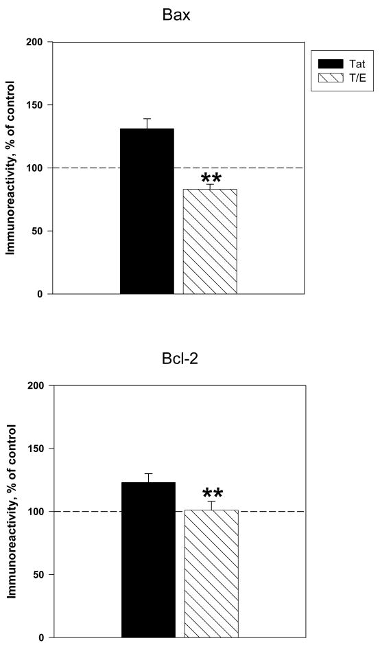
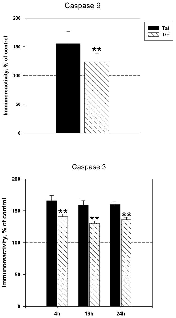
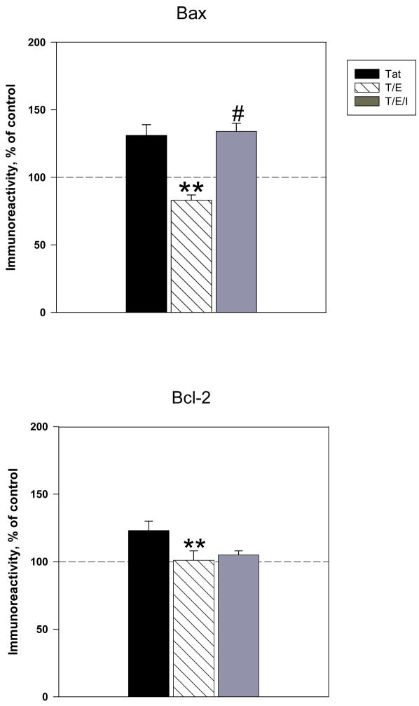
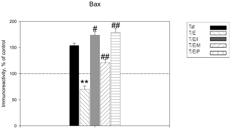
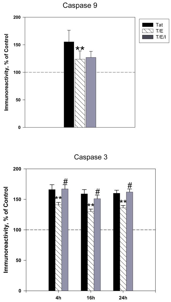
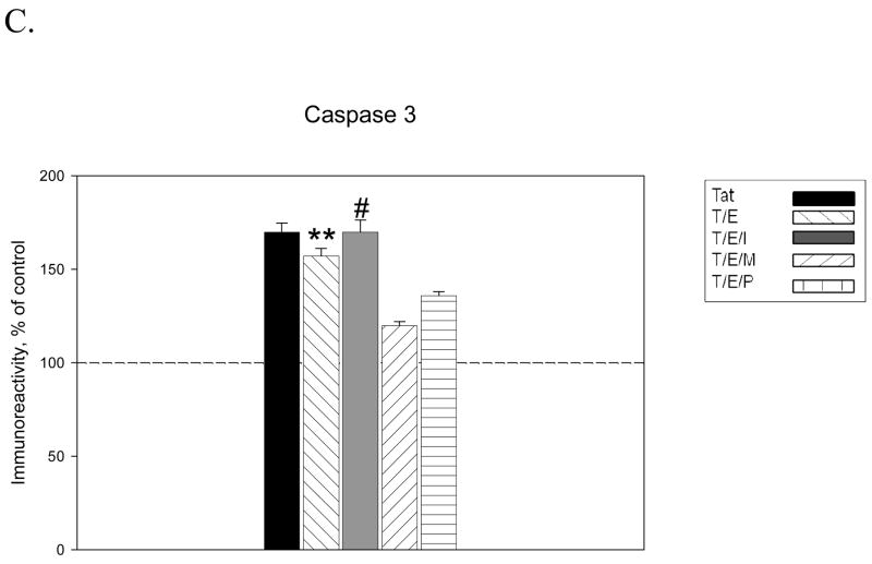
The protective actions of estrogen have been well evaluated in various models of neurodegeneration. These neuroprotective mechanisms may include a direct neuronal antiapoptotic effect as estrogen modulates actions of key regulators of the mitochondrial/intrinsic apoptotic cascade. We tested the ability of estrogen to protect against apoptotic signaling in cortical cell cultures exposed to Tat 1-86 (50 nM), and additionally, whether the beneficial actions of estrogen involved an estrogen receptor sensitive mechanism. We demonstrated that estrogen pretreatment significantly delayed Tat-induced cell death in primary cortical cultures. Pretreatment with 17β-estradiol (10 nM) attenuated the increased expression of antiapoptotic protein Bcl-2, proapoptotic protein Bax and activation of caspases linked to mitochondrial apoptotic pathway following Tat exposure. In addition, select components of apoptotic pathway signaling appear more sensitive to estrogen receptor (ER) activation, as the addition of ER antagonist ICI 182780 reversed estrogen downregulation of Bax and caspase 3, while estrogen effects on Tat-induced Bcl-2 and caspase 9 expression were maintained. Moreover, the addition of preferential ERα and ERβ antagonists (MPP dihydrochloride and PHTPP) indicated that estrogen effects on caspase 3 may be mediated by both receptor subtypes, whereas, was more involved in estrogen effects on Bax. Our data suggest that estrogen intervenes against HIV-1 Tat-induced cortical neuronal dysfunction via intersecting mitochondrial apoptotic pathway signaling in an ER-sensitive manner.


Figures

Figure 1
Figure 1. Estrogen treatment delays HIV-1 Tat 1-86 mediated decrease of cell viability in primary rat cortical cultures
The graph demonstrates the decrease in live cells in primary cultures following exposure to 50 nM Tat 1-86. Addition of 10nM 17β-estradiol 24h prior to Tat 1-86 exposure was able to significantly delay onset of cell death in primary neuronal cultures. Data presented as mean values, n of sister cultures analyzed 5–10 per each time point. *P<0.05 as compared to non-treated controls, #P<0.05 as compared to Tat-treated vs. Tat+E treated cultures
Figure 2
Figure 2. Time course of apoptotic protein expression following Tat exposure
Cortical lysates were collected at 4h, 16h and 24h following Tat 1-86 (50nM) exposure. Expression of Bcl-2, Bax, active Caspase 9 and active Caspase 3 were assessed by ELISA experiments. Results are presented as % of control values, experiments performed in triplicate, *P< 0.05 as compared to controls
Figure 3
Figure 3. 17β-estradiol attenuates Tat-induced expression of Bcl-2 and Bax protein levels
Cortical cultures were treated with 10nM 17β-estradiol 24h prior to Tat exposure. Expression of apoptotic proteins A. Bax (4h Tat exposure) and B. Bcl-2 (16h of Tat exposure) were assessed by ELISA experiments. Results are presented as % of control value, with experiments performed in triplicate, *P< 0.05 as compared to Tat-treated cultures. Legend box: Tat (T), Tat+Estrogen (T/E). Reference line in graph represents control group.
Figure 4
Figure 4. 17β-estradiol attenuates Tat-induced increases in active caspase expression
Cortical cultures were treated with 10nM 17β-estradiol 24h prior to Tat exposure. Expression of active A. Caspase 9 and B. Caspase 3 were assessed by ELISA experiments. Results are presented as % of control value, with experiments performed in triplicate, *P< 0.05 as compared to Tat-treated cultures. Legend box: Tat (T), Tat+Estrogen (T/E). Reference line in graph represents control group.
Figure 5
Figure 5. Tat-induced Bax expression sensitive to estrogen receptor -mediated estrogenic actions
A. Estrogen receptor antagonist, ICI 182,780, was added to cultures 1h before treatment with 17β-estradiol. Estrogen effects on Tat-induced increased expression of Bax were reversed by ICI 182,780, suggesting that estrogenic actions on this protein are mediated by ER signaling. B. Estrogen effects on Bcl-2 expression were not attenuated by the addition of ER antagonist. C. Specific antagonists for ERα (MPP, 100nM) and ERβ (PHTPP, 100nM) were added to cultures 30 min before treatment with 17β-estradiol. ER subtype specific antagonists reveal that ERβ mediated signaling preferential for estrogen effects on Bax. Experiments performed in triplicate, *P<0.05 as compared to Tat-treated cultures, **P<0.05 compared to Tat+E treated cultures. Legend box: Tat (T), Tat+Estrogen (T/E), T/E+ICI182, 780 (T/E/I), T/E+ MPP (T/E/M), T/E+PHTPP (T/E/P). Reference line in graph represents control group.
Figure 6
Figure 6. Estrogen receptor mediates estrogen effects on Tat-induced Caspase 3 not Caspase 9 active expression
Estrogen receptor antagonist, ICI 182,780, was added to cultures 1h before treatment with 17β-estradiol. A. Estrogen effects on caspase 9 expression were maintained in presence of ER antagonist. B. Estrogen effects on Tat-induced expression of caspase 3 were reversed by ICI 182,780, suggesting that estrogenic actions on caspase 3 are ER mediated. C. Estrogen effects against Tat-induced caspase 3 expression were maintained in presence of specific antagonists for ERα (MPP, 100nM) and ERβ (PHTPP, 100nM). Experiments performed in triplicate, *P<0.05 as compared to Tat-treated cultures, **P<0.05 as compared to Tat+E treated cultures. Legend box: Tat (T), Tat+Estrogen (T/E), T/E+ICI182780 (T/E/I), T/E+ MPP (T/E/M), T/E+PHTPP (T/E/P). Reference line in graph represents control group.

References

    1. Aksenov MY, Hasselrot U, Bansal Ak, et al. Oxidative damage induced by the injection of HIV-1 Tat protein in the rat striatum. Neurosci Lett. 2001;305:5–8. - PubMed
    1. Aksenov MY, Hasselrot U, Wu G, et al. Temporal relationships between HIV-1 Tat-induced neuronal degeneration, OX-42 immunoreactivity, reactive astrocytosis, and protein oxidation in the rat striatum. Brain Res. 2003;987:1–9. - PubMed
    1. Aksenova MV, Aksenov MY, Adams SM, Mactutus CF, Booze RM. Neuronal survival and resistance to HIV-1 Tat toxicity in the primary culture of rat fetal neurons. Exp Neurol. 2009;5(2):253–263. - PMC - PubMed
    1. Amantea D, Russo R, Bagetta G, Corasaniti MT. From clinical evidence to molecular mechanisms underlying neuroprotection afforded by estrogens. Pharmacol Res. 2005;52:119–132. - PubMed
    1. Ances BM, Ellis RJ. Dementia and neurocognitive disorders due to HIV-1 infection. Semin Neurol. 2007;27:86–92. - PubMed

Publication types

MeSH terms