Review
. 2010:33:245-67.
doi: 10.1146/annurev-neuro-060909-153248.

Wallerian degeneration, wld(s), and nmnat

Affiliations
Review

Wallerian degeneration, wld(s), and nmnat

Michael P Coleman et al. Annu Rev Neurosci. 2010.

Abstract

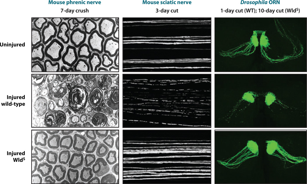
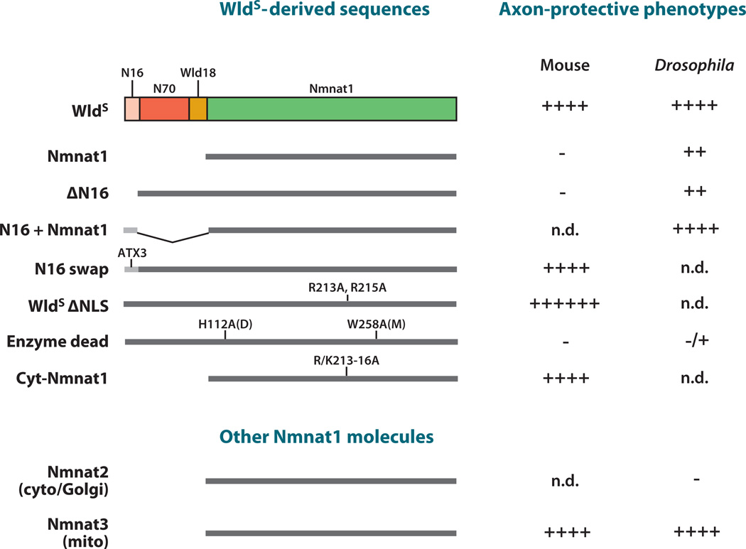
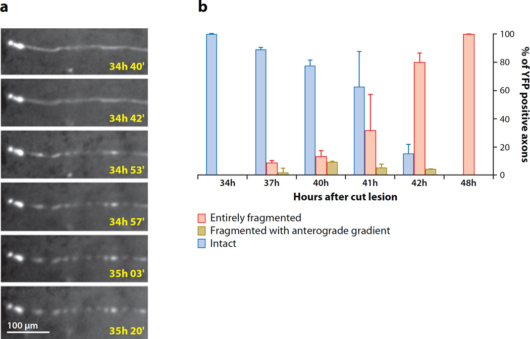
Traditionally, researchers have believed that axons are highly dependent on their cell bodies for long-term survival. However, recent studies point to the existence of axon-autonomous mechanism(s) that regulate rapid axon degeneration after axotomy. Here, we review the cellular and molecular events that underlie this process, termed Wallerian degeneration. We describe the biphasic nature of axon degeneration after axotomy and our current understanding of how Wld(S)--an extraordinary protein formed by fusing a Ube4b sequence to Nmnat1--acts to protect severed axons. Interestingly, the neuroprotective effects of Wld(S) span all species tested, which suggests that there is an ancient, Wld(S)-sensitive axon destruction program. Recent studies with Wld(S) also reveal that Wallerian degeneration is genetically related to several dying back axonopathies, thus arguing that Wallerian degeneration can serve as a useful model to understand, and potentially treat, axon degeneration in diverse traumatic or disease contexts.


Figures

Figure 1
Figure 1
Wallerian degeneration in wild-type axons and preservation in WldS. Injured wild-type axons (middle row) exhibit granular disintegration of the cytoskeleton seen in electron microscopy (left column) and fragmentation visualized by fluorescence microscopy (middle and right columns). Cytoskeletal integrity, unswollen mitochondria, and axon continuity are preserved by the WldS gene in each case (bottom row). Note the remarkable consistency of Wallerian degeneration and the neuroprotective WldS phenotype between mice and Drosophila. ORN, olfactory receptor neuron. Left column from Brown et al. (1994); reprinted with permission from Wiley-Blackwell. Middle column from Conforti et al. (2007b); reprinted by permission from Macmillan Publishers Ltd., Nature Publishing Group.
Figure 2
Figure 2
Axon protection mediated by WldS, WldS domains, and Nmnat molecules. The in vivo protective effects have been studied extensively in mouse and Drosophila. (Top) WldS-derived sequences represent constructs where specific domains of WldS were deleted or mutated. (Bottom) Other Nmnat molecules are mouse Nmnat2 and Nmnat3. Protection in either mouse or Drosophila is shown to the right. Protection and its relative strength are indicated by (+), a lack of protection is indicated by (−), and those not determined in vivo are indicated by n.d. Point mutations or domain swaps are shown above the diagram of each molecule, with point mutation positions that refer to their relative position in WldS.
Figure 3
Figure 3
Wallerian degeneration in wild-type axons is a biphasic process. (a) Distal stumps of injured wild-type axons in the dorsal column of mouse spinal cord remain continuous for a latent phase of over 34 h before fragmenting over the course of a few minutes. From Kerschensteiner et al. (2005) reprinted by permission from Macmillan Publishers Ltd., Nature Publishing Group. (b) Quantification of fragmented, unfragmented, and partially fragmented wild-type axons in distal sciatic nerve after transection injury shows a similar latent phase followed by fragmentation of all axons in the nerve over the next few hours. The timing of fragmentation is heterogenous within the axon population, but once started, fragmentation is rapid, such that the percentage of partially fragmented axons never exceeds 10% at any one time. From Beirowski et al. (2005) reprinted with permission from BioMed Central.

Similar articles

Cited by

References

    1. Adalbert R, Gillingwater TH, Haley JE, Bridge K, Beirowski B, et al. A rat model of slow Wallerian degeneration (WldS) with improved preservation of neuromuscular synapses. Eur. J. Neurosci. 2005;21:271–277. - PubMed
    1. Adalbert R, Nogradi A, Babetto E, Janeckova L, Walker SA, et al. Severely dystrophic axons at amyloid plaques remain continuous and connected to viable cell bodies. Brain. 2009;132:402–416. - PubMed
    1. Adalbert R, Nogradi A, Szabo A, Coleman MP. The slow Wallerian degeneration gene in vivo protects motor axons but not their cell bodies after avulsion and neonatal axotomy. Eur. J. Neurosci. 2006;24:2163–2168. - PubMed
    1. Alvarez S, Moldovan M, Krarup C. Acute energy restriction triggers Wallerian degeneration in mouse. Exp. Neurol. 2008;212:166–178. - PubMed
    1. Araki T, Sasaki Y, Milbrandt J. Increased nuclear NAD biosynthesis and SIRT1 activation prevent axonal degeneration. Science. 2004;305:1010–1013. - PubMed

Publication types

MeSH terms

LinkOut - more resources