Review
. 2010 May;7(5):577-87.
doi: 10.1517/17425240903571614.

Targeting gold nanocages to cancer cells for photothermal destruction and drug delivery

Affiliations
Review

Targeting gold nanocages to cancer cells for photothermal destruction and drug delivery

Claire M Cobley et al. Expert Opin Drug Deliv. 2010 May.

Abstract

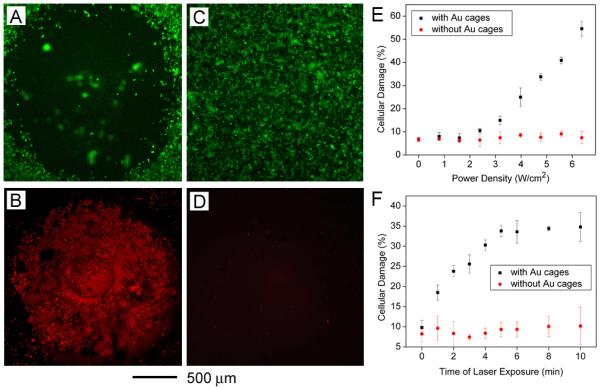
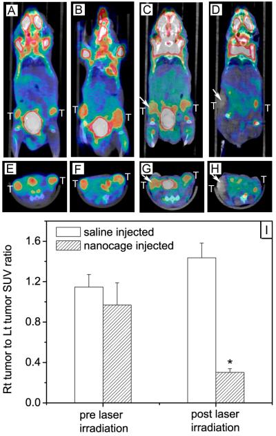
Importance of the field: Plasmonic nanoparticles provide a new route to treat cancer owing to their ability to convert light into heat effectively for photothermal destruction. Combined with the targeting mechanisms possible with nanoscale materials, this technique has the potential to enable highly targeted therapies to minimize undesirable side effects.

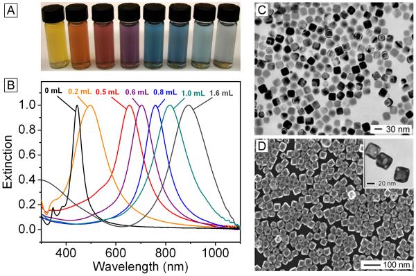
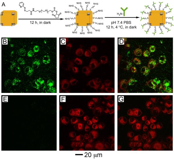
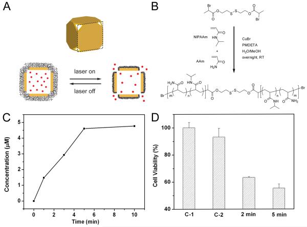
Areas covered in this review: This review discusses the use of gold nanocages, a new class of plasmonic nanoparticles, for photothermal applications. Gold nanocages are hollow, porous structures with compact sizes and precisely controlled plasmonic properties and surface chemistry. Also, a recent study of gold nanocages as drug-release carriers by externally controlling the opening and closing of the pores with a smart polymer whose conformation changes at a specific temperature is discussed. Release of the contents can be initiated remotely through near-infrared irradiation. Together, these topics cover the years from 2002 to 2009.

What the reader will gain: The reader will be exposed to different aspects of gold nanocages, including synthesis, surface modification, in vitro studies, initial in vivo data and perspectives on future studies.

Take home message: Gold nanocages are a promising platform for cancer therapy in terms of both photothermal destruction and drug delivery.


Figures

Figure 1
Figure 1
(A) Photograph and (B) UV-Vis spectra of gold nanocages tuned to a range of specific wavelengths by controlling the amount (volume listed above each trace) of chloroauric acid added into the reaction system. Modified with permission from [26], Copyright 2007 Nature Publishing Group. (C) Transmission electron microscopy (TEM) image of silver nanocubes, the starting template for the synthesis of gold nanocages. Modified with permission from [17], Copyright 2006 Royal Society of Chemistry. (D) Scanning electron microscopy (SEM) image of the gold nanocages, with TEM inset. Modified with permission from [44], Copyright 2005 Wiley-VCH.
Figure 2
Figure 2
(A) Schematic illustrating the protocol for conjugating antibodies to the surface of nanocages to create immuno gold nanocages. (B-D) U87MGwtEGFR cells after incubation with gold nanocages functionalized with EGFR antibodies. (E-G) Cells after incubation with gold nanocages covered by PEG. (B, E) Two-photon images of gold nanocages showing little uptake for the PEG-covered nanocages. (C, F) Fluorescence images of SK-BR-3 cells with FM4-64 dye used to stain the membranes. (D, G) Overlay of the images from gold nanocages and from FM4-64 dyes, showing clear overlap between the two for the EGFR-covered nanocages. Modified with permission from [51], Copyright 2009 American Chemical Society.
Figure 3
Figure 3
Qualitative and quantitative analysis of the photothermal effect of gold nanocages in vitro. (A, B) The cells were incubated with gold nanocages and illuminated for 5 min at a power density of 1.5 W/cm2 and examined with a fluorescence microscope. (C, D) Control experiments with the same laser irradiation, but no nanocage present. (A, C) Calcein AM assay, where green fluorescence indicates live cells. (B, D) ethidium homodimer 1 assay, where red fluorescence indicates dead cells. Modified with permission from [30], Copyright 2007 American Chemical Society. (E) Dependence of cellular damage on laser power density (irradiation time: 5 min). Data from cells incubated with immuno gold nanocages is depicted in black while data from controls with no gold nanocages is depicted in red. Though only 9.8% of the cells in the plate were in the irradiated area, the heat spread to surrounding areas to increase cellular damage. (F) Dependence of cellular damage on laser exposure time (power density: 4.77 W/cm2). Modified with permission from [59], Copyright 2008 American Chemical Society.
Figure 4
Figure 4
18F-FDG PET/CT co-registered images (coronal plane: A-D; axial plane: E-H) of mice injected with either gold nanocages or saline solution before and after laser irradiation: (A, E) a saline-injected mouse prior to laser irradiation; (B, F) a nanocage-injected mouse prior to laser irradiation; (C, G) the saline-injected mouse after laser irradiation; and (D, H) the nanocage-injected mouse after laser irradiation. The white arrows indicated the tumors that were exposed to the diode laser at a power density of 0.7 W/cm2 for 10 min. (I) Plot showing the ratios of laser-treated tumor to non-treated tumor for 18F-FDG standardized uptake values (SUV, P<0.001) [66], Copyright 2010 Wiley-VCH.
Figure 5
Figure 5
(A) Schematic illustrating the release mechanism for gold nanocages coated by smart polymer chains. (B) Atom transfer radical polymerization of NIPAAm and AAm monomers (at a molar ratio of m/n) as initiated by a disulfide initiator and in the presence of a Cu(I) catalyst. (C) Concentration of Dox released by heating the system at 45 °C for different periods of time. (D) Cell viability after pulsed laser irradiation at 20 mW/cm2 for 2 and 5 min, respectively, in the presence of Dox-loaded nanocages. Controls: no nanocages present (C-1) and in the presence of Dox-free nanocages (C-2), both irradiated for 2 min. Modified with permission from [46], Copyright 2009 Nature Publishing Group.

References

    1. Riehemann K, Schneider SW, Luger TA, et al. Nanomedicine - challenge and perspectives. Angew Chem Int Ed. 2009;48:872–897. ** Broad overview of recent advances and future perspectives in the field of nanomedicine.

    1. Heath JR, Davis ME. Nanotechnology and cancer. Annu Rev Med. 2008;59:251–653. - PMC - PubMed
    1. Peer D, Karp JM, Hong S, et al. Nanocarriers as an emerging platform for cancer therapy. Nat Nanotech. 2007;2:751–60. ** Broad overview of ways nanotechnology can be used for cancer treatment, including the current development stage of different therapies.

    1. Ferrari M. Cancer nanotechnology: Opportunities and challenges. Nat Rev Cancer. 2005;5:161–171. - PubMed
    1. Arap W, Pasqualini R, Ruoslahti E. Cancer treatment by targeted drug delivery to tumor vasculature in a mouse model. Science. 1998;279:377–80. - PubMed

Publication types