. 2010 Jun;113(6):1504-15.
doi: 10.1111/j.1471-4159.2010.06694.x. Epub 2010 Mar 24.

Serotonin 5-HT2C receptor protein expression is enriched in synaptosomal and post-synaptic compartments of rat cortex

Affiliations

Serotonin 5-HT2C receptor protein expression is enriched in synaptosomal and post-synaptic compartments of rat cortex

Noelle C Anastasio et al. J Neurochem. 2010 Jun.

Abstract

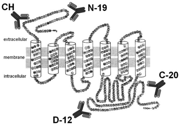
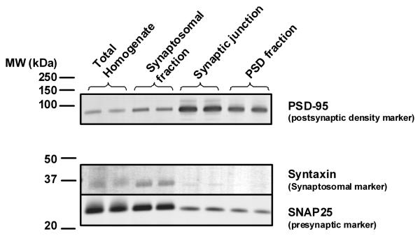
The action of serotonin (5-HT) at the 5-HT(2C) receptor (5-HT(2C)R) in cerebral cortex is emerging as a candidate modulator of neural processes that mediate core phenotypic facets of several psychiatric and neurological disorders. However, our understanding of the neurobiology of the cortical 5-HT(2C)R protein complex is currently limited. The goal of the present study was to explore the subcellular localization of the 5-HT(2C)R in synaptosomes and the post-synaptic density, an electron-dense thickening specialized for post-synaptic signaling and neuronal plasticity. Utilizing multiples tissues (brain, peripheral tissues), protein fractions (synaptosomal, post-synaptic density), and controls (peptide neutralization, 5-HT(2C)R stably-expressing cells), we established the selectivity of two commercially available 5-HT(2C)R antibodies and employed the antibodies in western blot and immunoprecipitation studies of prefrontal cortex (PFC) and motor cortex, two regions implicated in cognitive, emotional and motor dysfunction. For the first time, we demonstrated the expression of the 5-HT(2C)R in post-synaptic density-enriched fractions from both PFC and motor cortex. Co-immunoprecipitation studies revealed the presence of post-synaptic density-95 within the 5-HT(2C)R protein complex expressed in PFC and motor cortex. Taken together, these data support the hypothesis that the 5-HT(2C)R is localized within the post-synaptic thickening of synapses and is therefore positioned to directly modulate synaptic plasticity in cortical neurons.


Figures

Figure 1
Figure 1
Schematic representation of the 5-HT2CR topology and the target regions for the commercially available anti-5-HT2CR antibodies. Adapted from Julius et al. 1988a.
Figure 2
Figure 2. Specificity of anti-5-HT2CR antibodies for brain
Membrane fractions from motor cortex, PFC, cerebellum, liver, kidney and lung (n = 3-4 samples/each) were analyzed by Western blot. Densitometric analyses were conducted on the [A] D-12 (46 kDa), [B] N-19 (52 and 60 kDa), [C] C-20 (41 kDa) and [D] CH (41 and 60 kDa) IR bands. Results represent the mean density (± SEM) of the respective IR band normalized to β-actin. Data were analyzed using a one-way ANOVA followed by Dunnett's procedure. *p<0.05 vs. motor cortex for [A] D-12 46 kDa band, [B] N-19 52 kDa band, [C] C-20 41 kDa band and [D] CH 41 kDa band. ^p<0.05 vs. motor cortex for [B] N-19 60 kDa band.
Figure 3
Figure 3. Western blot analysis determining the detection limits of 5-HT2CR with the N-19 antibody in the synaptosomal fraction of PFC and motor cortex
Various concentrations (10, 15, and 20 μg) of synaptosomal protein from the PFC [A and B] or motor cortex [C and D] were analyzed by Western blot using the N-19 antibody. The N-19 antibody detected multiple bands of various intensities, including the previously described 52 and 60 kDa bands in both PFC [A] and motor cortex [C]. Peptide neutralization of the N-19 antibody with its appropriate blocking peptide greatly reduced the intensity of the 52 and 60 kDa IR bands in both PFC [B] and motor cortex [D].
Figure 4
Figure 4. Western blot analysis showing the specificity of 5-HT2CR antibodies for the 5-HT2CR in CHO cells
Crude membrane protein from parental CHO cells, which do not natively express the 5-HT2CR, or CHO cells stably expressing the human 5-HT2CR (1C19) were analyzed by Western blot analysis using the D-12 [A] or N-19 [B] antibody. The D-12 antibody detected the 46 kDa band only in the 1C19 cell line [A]. The N-19 antibody detected the 52 kDa band specifically in the 1C19 cell line, while the 41 kDa band was detected in both the parental and 1C19 cell lines [B]. The 60 kDa band detected in rat brain preparations labeled by N-19 was not detected in the parental or 1C19 cell lines [B].
Figure 5
Figure 5. Immunoprecipitation studies with anti-5-HT2CR N-19 or D-12 antibody in PFC synaptosomes
Synaptosomal fractions were immunoprecipitated (IP) with D-12 antibody and immunoblotted (IB) with D-12 [A] or N-19 [B] antibody. The D-12 antibody detected an intense IR band at 46 kDa and a fainter band at 52 kDa following immunoprecipitation with D-12 [A]. The D-12 immunoprecipitation and subsequent immunoblotting with the N-19 antibody resulted in detection of two bands at 52 and 60 kDa [B]. Synaptosomal fractions were immunoprecipitated with N-19 antibody and immunoblotted with D-12 [C] or N-19 [D] antibody. The D-12 antibody detected a single band at 46 kDa following immunoprecipitation with N-19 [C]. N-19 immunoprecipitation and subsequent immunoblotting with the N-19 antibody detected two bands at 52 and 60 kDa [D].
Figure 6
Figure 6. Postsynaptic localization of the 5-HT2CR in PFC
Cortical synaptosomal fractions were immunoprecipitated with D-12 [A] or N-19 [B] and immunoblotted with PSD-95 antibody. The PSD-95 antibody detected the expected IR band at 95 kDa and a faint band at 100 kDa in PFC synaptosomal samples immunoprecipitated with the D-12 [A]. The PSD-95 antibody detected a band at 95 kDa in synaptosomes immunoprecipitated with the N-19 antibody [B].
Figure 7
Figure 7. Validation of synaptosomal enrichment from motor cortex
Total homogenate, synaptosomal fraction, synaptic junction and postsynaptic density-enriched fraction isolated from motor cortex were analyzed by Western blot using antibodies against PSD-95 (postsynaptic density marker), syntaxin (presynaptic active zone marker) and SNAP-25 (presynpatic active zone marker).
Figure 8
Figure 8. Band patterns of 5-HT2CR in the postsynaptic density from motor cortex
PSD-enriched fractions from motor cortex were analyzed by Western blot using antibodies against the 5-HT2CR Lane 1: D-12 immunoblot shows an IR band at MW of 46 kDa. Lane 2: N-19 immunoblot shows an IR bands at MWs of 52 and 60 kDa as well as the nonspecific 41 kDa band.

References

    1. Abbas AI, Yadav PN, Yao WD, Arbuckle MI, Grant SG, Caron MG, Roth BL. PSD-95 is essential for hallucinogen and atypical antipsychotic drug actions at serotonin receptors. J. Neurosci. 2009;29:7124–7136. - PMC - PubMed
    1. Abramowski D, Rigo M, Duc D, Hoyer D, Staufenbiel M. Localization of the 5-hydroxytryptamine2C receptor protein in human and rat brain using specific antisera. Neuropharmacology. 1995;34:1635–1645. - PubMed
    1. Abramowski D, Staufenbiel M. Identification of the 5-hydroxytryptamine2C receptor as a 60-kDa N-glycosylated protein in choroid plexus and hippocampus. J. Neurochem. 1995;65:782–790. - PubMed
    1. Backstrom JR, Westphal RS, Canton H, Sanders-Bush E. Identification of rat serotonin 5-HT2C receptors as glycoproteins containing N-linked oligosaccharides. Brain Res. Mol. Brain Res. 1995;33:311–318. - PubMed
    1. Banker G, Churchill L, Cotman CW. Proteins of the postsynaptic density. J Cell Biol. 1974;63:456–465. - PMC - PubMed

Publication types

Substances