

Collagen fibril morphology and mechanical properties of the Achilles tendon in two inbred mouse strains
- PMID: 20345854
- PMCID: PMC2952385
- DOI: 10.1111/j.1469-7580.2010.01225.x
Collagen fibril morphology and mechanical properties of the Achilles tendon in two inbred mouse strains
Abstract
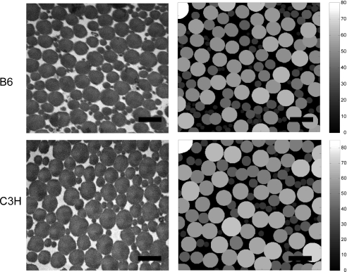
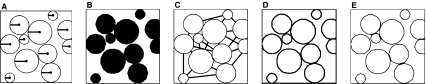
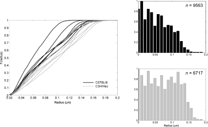
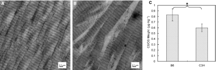
The relationship between collagen fibril morphology and the functional behavior of tendon tissue has been investigated in numerous experimental studies. Several of these studies suggest that larger fibril radius is a primary determinant of higher tendon stiffness and strength; others have shown that factors apart from fibril radius (such as fibril-fibril interactions) may be critical to improved tendon strength. In the present study, we investigate these factors in two inbred mouse strains that are widely used in skeletal structure-function research: C57BL/6J (B6) and C3H/HeJ (C3H). The aim was to establish a quantitative baseline that will allow one to assess how regulation of tendon extracellular matrix architecture affects tensile mechanical properties. We specifically focused on collagen fibril structure and glycosaminoglycan (GAG) content--the two primary constituents of tendon by dry weight--and their potential functional interactions. For this purpose, Achilles tendons from both groups were tested to failure in tension. Tendon collagen morphology was analyzed from transmission electron microscopy images of tendon sections perpendicular to the longitudinal axis. Our results showed that the two inbred strains are macroscopically similar, but C3H mice have a higher elastic modulus (P < 0.05). Structurally, C3H mice showed a larger collagen fibril radius compared to B6 mice (96 +/- 7 nm and 80 +/- 10 nm respectively). Tendons from C3H mice also showed smaller specific fibril surface (0.015 +/- 0.001 nm nm(-2) vs. 0.017 +/- 0.003 nm nm(-2) in the B6 tendons, P < 0.05), and accordingly a lower concentration of GAGs (0.60 +/- 0.07 microg mg(-1) vs. 0.83 +/- 0.11 microg mg(-1), P < 0.05). As in other studies of tendon structure and function, larger collagen fibril radius appears to be associated with stiffer tendon, but this functional difference could also be attributed to reduced potential surface area exchange between fibrils and the surrounding proteoglycan-rich matrix, in which the hydrophilic GAG side chains may promote inter-fibril sliding. This study provides an architectural and functional baseline for a comparative murine model that can be used to investigate the genetic regulation of tendon biomechanics.
Figures
Similar articles
-
Tendon glycosaminoglycan proteoglycan sidechains promote collagen fibril sliding-AFM observations at the nanoscale.J Biomech. 2013 Feb 22;46(4):813-8. doi: 10.1016/j.jbiomech.2012.11.017. Epub 2012 Dec 6. J Biomech. 2013. PMID: 23219277
-
Equivalent stiffness after glycosaminoglycan depletion in tendon--an ultra-structural finite element model and corresponding experiments.J Theor Biol. 2011 Jan 7;268(1):77-83. doi: 10.1016/j.jtbi.2010.10.007. Epub 2010 Oct 13. J Theor Biol. 2011. PMID: 20950629
-
Ultrastructural determinants of murine achilles tendon strength during healing.Connect Tissue Res. 2003;44(5):218-24. Connect Tissue Res. 2003. PMID: 14660092
-
Collagen structure of tendon relates to function.ScientificWorldJournal. 2007 Mar 30;7:404-20. doi: 10.1100/tsw.2007.92. ScientificWorldJournal. 2007. PMID: 17450305 Free PMC article. Review.
-
Structure of the tendon connective tissue.Scand J Med Sci Sports. 2000 Dec;10(6):312-20. doi: 10.1034/j.1600-0838.2000.010006312.x. Scand J Med Sci Sports. 2000. PMID: 11085557 Review.
Cited by
-
Turnover of fibrillar collagen in soft biological tissue with application to the expansion of abdominal aortic aneurysms.J R Soc Interface. 2012 Dec 7;9(77):3366-77. doi: 10.1098/rsif.2012.0416. Epub 2012 Aug 15. J R Soc Interface. 2012. PMID: 22896562 Free PMC article.
-
Changing material properties of the tree shrew sclera during minus lens compensation and recovery.Invest Ophthalmol Vis Sci. 2015 Mar 3;56(3):2065-78. doi: 10.1167/iovs.14-15352. Invest Ophthalmol Vis Sci. 2015. PMID: 25736788 Free PMC article.
-
Functional tissue engineering of tendon: Establishing biological success criteria for improving tendon repair.J Biomech. 2014 Jun 27;47(9):1941-8. doi: 10.1016/j.jbiomech.2013.10.023. Epub 2013 Oct 22. J Biomech. 2014. PMID: 24200342 Free PMC article. Review.
-
Biomechanical properties and histology of db/db diabetic mouse Achilles tendon.Muscles Ligaments Tendons J. 2014 Nov 17;4(3):280-4. eCollection 2014 Jul. Muscles Ligaments Tendons J. 2014. PMID: 25489543 Free PMC article.
-
A quantitative study of the relationship between the distribution of different types of collagen and the mechanical behavior of rabbit medial collateral ligaments.PLoS One. 2014 Jul 25;9(7):e103363. doi: 10.1371/journal.pone.0103363. eCollection 2014. PLoS One. 2014. PMID: 25062068 Free PMC article.
References
-
- Akhter MP, Cullen DM, Pedersen EA, et al. Bone response to in vivo mechanical loading in two breeds of mice. Calcif Tissue Int. 1998;63:442–449. - PubMed
-
- Ault HK, Hoffman AH. A composite micromechanical model for connective tissues: part I – Theory. J Biomech Eng. 1992a;114:137–141. - PubMed
-
- Ault HK, Hoffman AH. A composite micromechanical model for connective tissues: part II – Application to rat tail tendon and joint capsule. J Biomech Eng. 1992b;114:142–146. - PubMed
-
- Battaglia TC, Clark RT, Chhabra A, et al. Ultrastructural determinants of murine achilles tendon strength during healing. Connect Tissue Res. 2003;44:218–224. - PubMed
Publication types
MeSH terms
Substances
LinkOut - more resources
Full Text Sources
Research Materials
