

Validation of the murine aortic arch as a model to study human vascular diseases
- PMID: 20345858
- PMCID: PMC2871992
- DOI: 10.1111/j.1469-7580.2010.01220.x
Validation of the murine aortic arch as a model to study human vascular diseases
Abstract
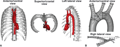
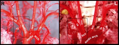
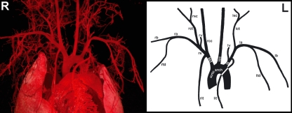
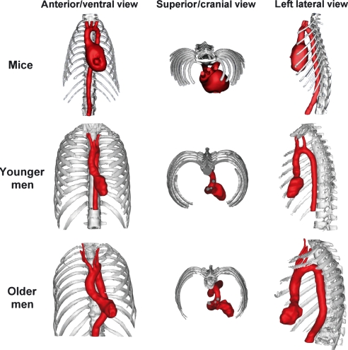
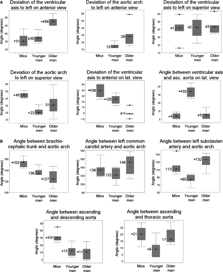
Although the murine thoracic aorta and its main branches are widely studied to gain more insight into the pathogenesis of human vascular diseases, detailed anatomical data on the murine aorta are sparse. Moreover, comparative studies between mice and men focusing on the topography and geometry of the heart and aorta are lacking. As this hampers the validation of murine vascular models, the branching pattern of the murine thoracic aorta was examined in 30 vascular corrosion casts. On six casts the intrathoracic position of the heart was compared with that of six younger and six older men of whom contrast-enhanced computer tomography images of the thorax were three-dimensionally reconstructed. In addition, the geometry of the human thoracic aorta was compared with that of the mouse by reconstructing micro-computer tomography images of six murine casts. It was found that the right brachiocephalic trunk, left common carotid artery and left subclavian artery branched subsequently from the aortic arch in both mice and men. The geometry of the branches of the murine aortic arch was quite similar to that of men. In both species the initial segment of the aorta, comprising the ascending aorta, aortic arch and cranial/superior part of the descending aorta, was sigmoidally curved on a cranial/superior view. Although some analogy between the intrathoracic position of the murine and human heart was observed, the murine heart manifestly deviated more ventrally. The major conclusion of this study is that, in both mice and men, the ascending and descending aorta do not lie in a single vertical plane (non-planar aortic geometry). This contrasts clearly with most domestic mammals in which a planar aortic pattern is present. As the vascular branching pattern of the aortic arch is also similar in mice and men, the murine model seems valuable to study human vascular diseases.
Figures
Similar articles
-
Aortic Arch and Thoracic Aorta Curvature Remodeling after Thoracic Endovascular Aortic Repair.Ann Vasc Surg. 2017 Jan;38:233-241. doi: 10.1016/j.avsg.2016.05.097. Epub 2016 Aug 12. Ann Vasc Surg. 2017. PMID: 27522975
-
Anatomical variations of the aortic arch branches in a sample of Chinese cadavers: embryological basis and literature review.Interact Cardiovasc Thorac Surg. 2019 Apr 1;28(4):622-628. doi: 10.1093/icvts/ivy296. Interact Cardiovasc Thorac Surg. 2019. PMID: 30445440 Review.
-
Anatomical variations of aortic arch branching: evaluation with computed tomographic angiography.Cardiol Young. 2014 Jun;24(3):485-93. doi: 10.1017/S1047951113000656. Epub 2013 May 22. Cardiol Young. 2014. PMID: 23694814 Review.
-
A systematic classification of the left-sided aortic arch variants based on cadaveric studies' prevalence.Surg Radiol Anat. 2021 Mar;43(3):327-345. doi: 10.1007/s00276-020-02625-1. Epub 2021 Jan 2. Surg Radiol Anat. 2021. PMID: 33386933
-
Extraordinary branching pattern of the aortic arch.Clin Anat. 2013 Nov;26(8):1006-7. doi: 10.1002/ca.22208. Epub 2013 Jan 27. Clin Anat. 2013. PMID: 23355323
Cited by
-
Casting techniques of equine hand and foot synovial cavities for the development of teaching models.Front Vet Sci. 2025 Mar 4;12:1524549. doi: 10.3389/fvets.2025.1524549. eCollection 2025. Front Vet Sci. 2025. PMID: 40104547 Free PMC article.
-
Performance comparison of ultrasound-based methods to assess aortic diameter and stiffness in normal and aneurysmal mice.PLoS One. 2015 May 29;10(5):e0129007. doi: 10.1371/journal.pone.0129007. eCollection 2015. PLoS One. 2015. PMID: 26023786 Free PMC article.
-
Recommendations for Design, Execution, and Reporting of Studies on Experimental Thoracic Aortopathy in Preclinical Models.Arterioscler Thromb Vasc Biol. 2025 May;45(5):609-631. doi: 10.1161/ATVBAHA.124.320259. Epub 2025 Mar 13. Arterioscler Thromb Vasc Biol. 2025. PMID: 40079138 Review.
-
Propagation-based phase-contrast synchrotron imaging of aortic dissection in mice: from individual elastic lamella to 3D analysis.Sci Rep. 2018 Feb 2;8(1):2223. doi: 10.1038/s41598-018-20673-x. Sci Rep. 2018. PMID: 29396472 Free PMC article.
-
A Method to Visualize and Quantify the Intraosseous Arteries of the Femoral Head by Vascular Corrosion Casting.Orthop Surg. 2022 Aug;14(8):1864-1872. doi: 10.1111/os.13319. Epub 2022 Jul 11. Orthop Surg. 2022. PMID: 35818638 Free PMC article.
References
-
- Anonymous . Terminologia Anatomica: International Anatomical Terminology. Stuttgart: Georg Thieme Verlag: Federative Committee on Anatomical Terminology (FCAT); 1998.
-
- Anonymous Report of the AVMA Panel on euthanasia. J Am Vet Med Assoc. 2001;218:669–696. - PubMed
-
- Anonymous . Nomina Anatomica Veterinaria. 5th ed. Hamburg, Columbia, Ghent, Sapporo: World Association of Veterinary Anatomists; 2005. Internet reference: http://www.wava-amav.org.
-
- Anonymous . Strasbourg: Council of Europe; 2006. Appendix A of the European convention for the protection of vertebrate animals used for experimental and other scientific purposes (ETS No. 123): Guidelines for accommodation and care of animals. 15 June 2006. Available at: http://conventions.coe.int.
Publication types
MeSH terms
LinkOut - more resources
Full Text Sources
Medical
Research Materials
