. 2010 May;216(5):589-99.
doi: 10.1111/j.1469-7580.2010.01221.x. Epub 2010 Mar 19.

Human midsagittal brain shape variation: patterns, allometry and integration

Affiliations

Human midsagittal brain shape variation: patterns, allometry and integration

Emiliano Bruner et al. J Anat. 2010 May.

Abstract

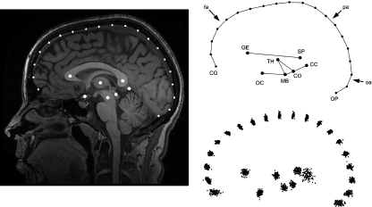
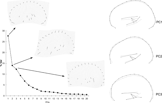
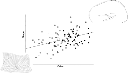
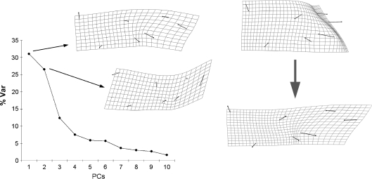
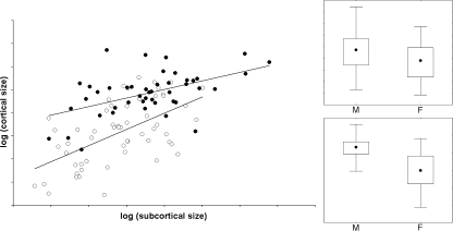
Midsagittal cerebral morphology provides a homologous geometrical reference for brain shape and cortical vs. subcortical spatial relationships. In this study, midsagittal brain shape variation is investigated in a sample of 102 humans, in order to describe and quantify the major patterns of correlation between morphological features, the effect of size and sex on general anatomy, and the degree of integration between different cortical and subcortical areas. The only evident pattern of covariation was associated with fronto-parietal cortical bulging. The allometric component was weak for the cortical profile, but more robust for the posterior subcortical areas. Apparent sex differences were evidenced in size but not in brain shape. Cortical and subcortical elements displayed scarcely integrated changes, suggesting a modular separation between these two areas. However, a certain correlation was found between posterior subcortical and parietal cortical variations. These results should be directly integrated with information ranging from functional craniology to wiring organization, and with hypotheses linking brain shape and the mechanical properties of neurons during morphogenesis.


Figures

Fig. 1
Fig. 1
The configuration is based onto seven subcortical landmarks and 20 cortical landmarks (fronto-parieto-occipital profile). The coordinates were superimposed using a Procrustes approach. The scatterplot (lower right) shows the variation of the whole sample after superimposition. CC, cerebro-cerebellar inner boundary; CG, crista galli; CO, colliculi; GE, genu; MB, midbrain; OC, optic chiasm; OP, internal occipital protuberance; SP, splenium; TH, centre of the thalamus. Arrows: frontal areas (fa), occipital areas (oa), parietal areas (pa).
Fig. 2
Fig. 2
Principal component analysis of the full configuration. The scree plot shows the distribution of the variance along the principal components, and the covariation patterns (thin plate spline deformation grids and wireframes) for the first three principal components.
Fig. 3
Fig. 3
Multivariate regression of shape (Procrustes coordinates) onto centroid size (males: black dots; females: white dots). The wireframe shows the pattern of covariation along the allometric vector. The deformation grid shows the reverse pattern (from larger to smaller size) after magnification, showing a crease at thalamic area.
Fig. 4
Fig. 4
Left: principal component analysis of the subcortical configuration. The scree plot shows the distribution of the variance along the principal components, and the covariation patterns (thin plate spline deformation grids) for the first two principal components. Right: allometric vector, from small (above, magnified) to large (below) size.
Fig. 5
Fig. 5
Left: log-log least-squares regression between subcortical and cortical size (males: black dots; females: white dots). Right: non-parametric distributions (median, interquartile, range) of the subcortical (above) and cortical (below) centroid size for males (M) and females (F).
Fig. 6
Fig. 6
(A) First latent vector from PLS regression between cortical (above) and subcortical (below) areas. (B) The partitions with lower correlation isolates posterior subcortical and parietal cortical landmarks from the rest of the configuration.

Similar articles

Cited by

References

    1. Ackermann RR, Cheverud JM. Detecting genetic drift versus selection in human evolution. Proc Natl Acad Sci U S A. 2004;101:17946–17951. - PMC - PubMed
    1. Bastir M. A systems-model for the morphological analysis of integration and modularity in human craniofacial evolution. J Anthropol Sci. 2008;86:37–58. - PubMed
    1. Bastir M, Rosas A. Mosaic evolution of the basicranium in Homo and its relation to modular development. Evol Biol. 2009;36:57–70.
    1. Bastir M, Rosas A, O’Higgins P. Craniofacial levels and the morphological maturation of the human skull. J Anat. 2006;209:637–654. - PMC - PubMed
    1. Battaglia-Mayer A, Caminiti R. Optic ataxia as a result of the breakdown of the global tuning fields of parietal neurons. Brain. 2002;125:225–237. - PubMed

Publication types

LinkOut - more resources