. 2010 Apr;277(8):1967-78.
doi: 10.1111/j.1742-4658.2010.07618.x. Epub 2010 Mar 18.

A new bright green-emitting fluorescent protein--engineered monomeric and dimeric forms

Affiliations

A new bright green-emitting fluorescent protein--engineered monomeric and dimeric forms

Robielyn P Ilagan et al. FEBS J. 2010 Apr.

Abstract

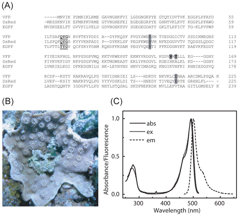
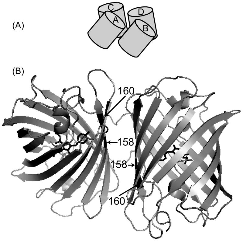
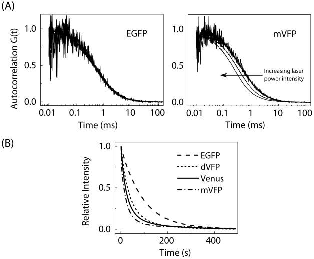
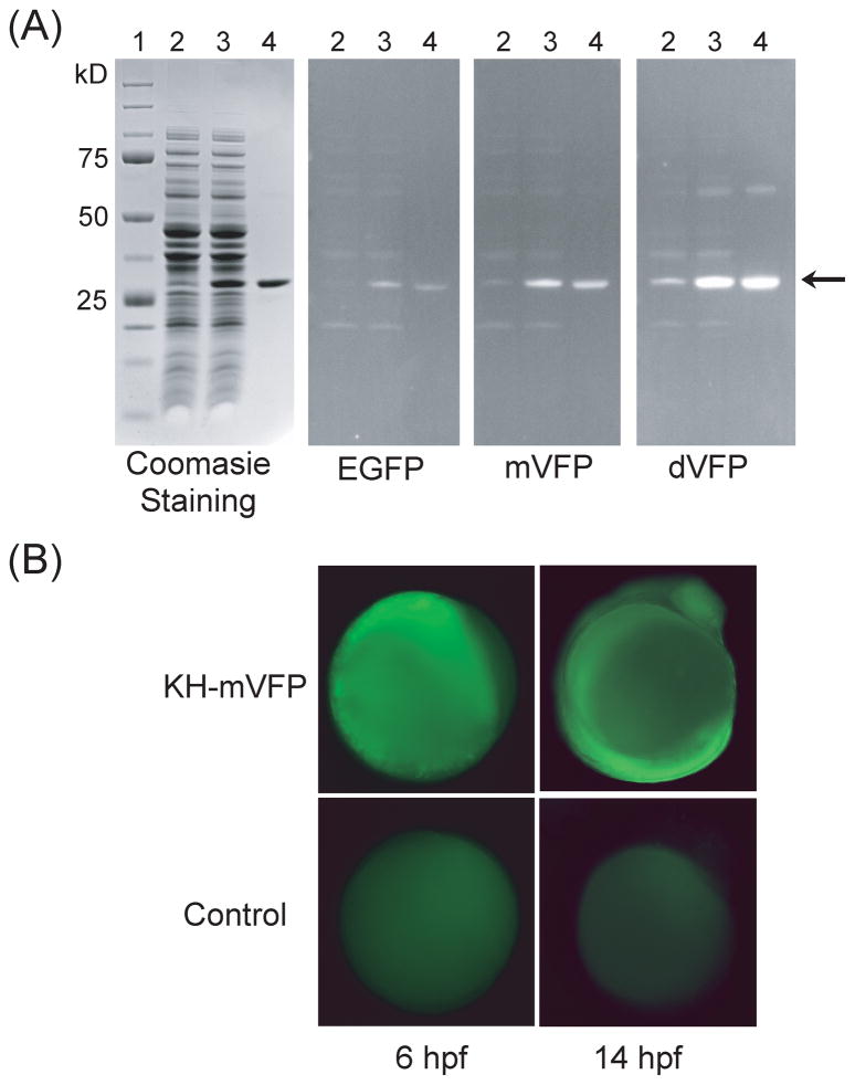
Fluorescent proteins have become essential tools in molecular and biological applications. Here, we present a novel fluorescent protein isolated from warm water coral, Cyphastrea microphthalma. The protein, which we named vivid Verde fluorescent protein (VFP), matures readily at 37 degrees C and emits bright green light. Further characterizations revealed that VFP has a tendency to form dimers. By creating a homology model of VFP, based on the structure of the red fluorescent protein, DsRed, we were able to make mutations that alter the protein's oligomerization state. We present two proteins, mVFP and mVFP1, that are both exclusively monomeric, and one protein, dVFP, which is dimeric. We characterized the spectroscopic properties of VFP and its variants in comparison with enhanced green fluorescent protein (EGFP), a widely used variant of GFP. All the VFP variants are at least twice as bright as EGFP. Finally, we demonstrated the effectiveness of the VFP variants in both in vitro and in vivo detection applications.


Figures

Figure 1
Figure 1
(A) Amino acid sequence alignment of VFP with DsRed and EGFP. The chromophore forming amino acid residues are highlighted in bold and underlined. Amino acid residues, N158 and T160 of VFP, where mutations were made are indicated by bold letter in gray background. The conserved Arg and Glu (corresponding to Arg96 and Glu222 of GFP) are highlighted in gray background. (B) A scleractinian coral, Cyphastrea microphthalma, collected in 1.2 m of water off Lizard Island on the Australian Great Barrier Reef. (C) Overlay of the absorption, fluorescence excitation and fluorescence emission spectra of VFP. The samples were excited at 450 nm and emission spectra were measured from 465 nm to 650 nm. Fluorescence excitation spectra were obtained from 250 nm to 515 nm by monitoring the emission at 530 nm. The spectra were normalized at the maximum peak.
Figure 2
Figure 2
(A) A cartoon illustration of DsRed tetramer arranged as a dimer of dimers with AB (=CD) and AC (=BD) interfaces. (B) Structure of two of the four subunits of the tetrameric DsRed consisting of the AC polar interface. The positions of amino acid residues at 158 and 160 (corresponding to 162 and 164, respectively, in DsRed) where mutations were made are indicated by arrows. The chromophore at the center of the β-barrel structure is shown in black sticks. Protein Data Bank (PDB) code: 1GGX. [55]
Figure 3
Figure 3
(A) Representative FCS autocorrelation curves of EGFP and mVFP taken at increasing laser power intensities from 0.25 to 5 μW. A shift in the autocorrelation curve to the left, to apparent shorter diffusion times, as a function of laser power intensity was observed for VFP and its variants. The autocorrelation curves are normalized to the number of molecules obtained from the fitting. (B) Photobleaching curves for the EGFP, Venus, mVFP and dVFP under mercury arc lamp illumination using wide-field microscope. The relative photostability of VFP and its variants are reported in Table 1.
Figure 4
Figure 4
(A) Comparison of the T-Mod fused to EGFP, mVFP, or dVFP as a replacement for antibodies in Western blot analysis. A duplicate SDS-PAGE gel used in Western blotting is stained with Coomassie Brilliant Blue. Lane (1) precision plus protein standard (BioRad); (2) lysate; (3) lysate supplemented with 1 mg/mL of purified GST-MEEVF protein; (4) purified GST-MEEVF. Arrow indicates the GST-MEEVF protein band. (B) Microinjection of KH-mVFP fusion mRNA into zebrafish embryos. The expression of KH-mVFP protein was monitored in the embryos at 6 hpf (hour post fertilization) and 14 hpf by fluorescence microscopy. Zebrafish embryos without RNA injections were used as a control.

References

    1. Shaner NC, Patterson GH, Davidson MW. Advances in fluorescent protein technology. J Cell Sci. 2007;120:4247–4260. - PubMed
    1. Day RN, Davidson MW. The fluorescent protein palette: tools for cellular imaging. Chem Soc Rev. 2009;38:2887–2921. - PMC - PubMed
    1. Lippincott-Schwartz J, Patterson GH. Photoactivatable fluorescent proteins for diffraction-limited and super-resolution imaging. Trends Cell Biol. 2009;19:555–565. - PMC - PubMed
    1. Nienhaus GU, Wiedenmann J. Structure, Dynamics and Optical Properties of Fluorescent Proteins: Perspectives for Marker Development. Chemphyschem. 2009;10:1369–1379. - PubMed
    1. Patterson GH, Lippincott-Schwartz J. A photoactivatable GFP for selective photolabeling of proteins and cells. Science. 2002;297:1873–1877. - PubMed

Publication types

Substances

Associated data