

Three lateral osteotomy designs for bilateral sagittal split osteotomy: biomechanical evaluation with three-dimensional finite element analysis
- PMID: 20346142
- PMCID: PMC2853503
- DOI: 10.1186/1746-160X-6-4
Three lateral osteotomy designs for bilateral sagittal split osteotomy: biomechanical evaluation with three-dimensional finite element analysis
Abstract
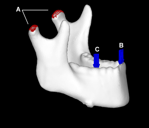
Background: The location of the lateral osteotomy cut during bilateral sagittal split osteotomy (BSSO) varies according to the surgeon's preference, and no consensus has been reached regarding the ideal location from the perspective of biomechanics. The purpose of this study was to evaluate the mechanical behavior of the mandible and screw-miniplate system among three lateral osteotomy designs for BSSO by using three-dimensional (3-D) finite element analysis (FEA).
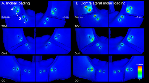
Methods: The Trauner-Obwegeser (TO), Obwegeser (Ob), and Obwegeser-Dal Pont (OD) methods were used for BSSO. In all the FEA simulations, the distal segments were advanced by 5 mm. Each model was fixed by using miniplates. These were applied at four different locations, including along Champy's lines, to give 12 different FEA miniplate fixation methods. We examined these models under two different loads.
Results: The magnitudes of tooth displacement, the maximum bone stress in the vicinity of the screws, and the maximum stress on the screw-miniplate system were less in the OD method than in the Ob and TO methods at all the miniplate locations. In addition, Champy's lines models were less than those at the other miniplate locations.
Conclusions: The OD method allows greater mechanical stability of the mandible than the other two techniques. Further, miniplates placed along Champy's lines provide greater mechanical advantage than those placed at other locations.
Figures


References
-
- Trauner R, Obwegeser H. The surgical correction of mandibular prognathism and retrognathia with consideration of genioplasty. I. Surgical procedures to correct mandibular prognathism and reshaping of the chin. Oral Surg Oral Med Oral Pathol. 1957;10:677–689. doi: 10.1016/S0030-4220(57)80063-2. contd. - DOI - PubMed
-
- Dal Pont G. Retromolar osteotomy for the correction of prognathism. J Oral Surg Anesth Hosp Dent Serv. 1961;19:42–47. - PubMed
-
- Hashiba Y, Ueki K, Marukawa K, Shimada M, Yoshida K, Shimizu C, Alam S, Nakagawa K. A comparison of lower lip hypoesthesia measured by trigeminal somatosensory-evoked potential between different types of mandibular osteotomies and fixation. Oral Surg Oral Med Oral Pathol Oral Radiol Endod. 2007;104:177–185. doi: 10.1016/j.tripleo.2006.11.038. - DOI - PubMed
-
- Cillo JE, Stella JP. Selection of sagittal split ramus osteotomy technique based on skeletal anatomy and planned distal segment movement: current therapy. J Oral Maxillofac Surg. 2005;63:109–114. - PubMed
Publication types
MeSH terms
LinkOut - more resources
Full Text Sources
Other Literature Sources
Miscellaneous
