

Proteome analysis of human Wharton's jelly cells during in vitro expansion
- PMID: 20346146
- PMCID: PMC2867805
- DOI: 10.1186/1477-5956-8-18
Proteome analysis of human Wharton's jelly cells during in vitro expansion
Abstract
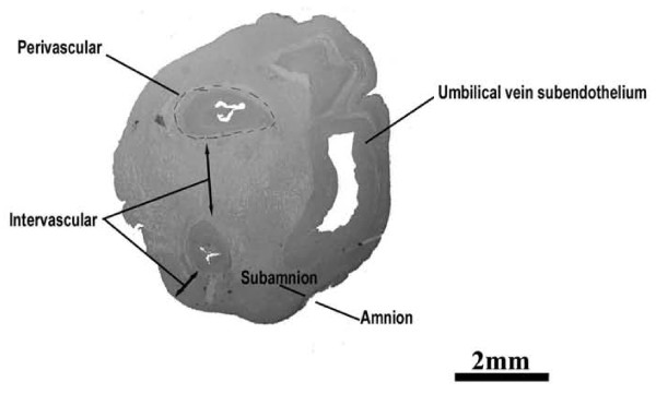
Background: The human umbilical cord contains mucoid connective tissue and fibroblast-like cells. These cells named Wharton's jelly cells, (WJCs) display properties similar to mesenchymal stem cells therefore representing a rich source of primitive cells to be potentially used in regenerative medicine.
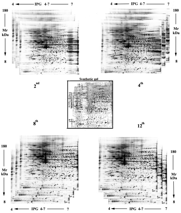
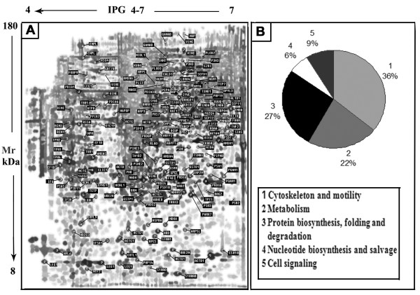
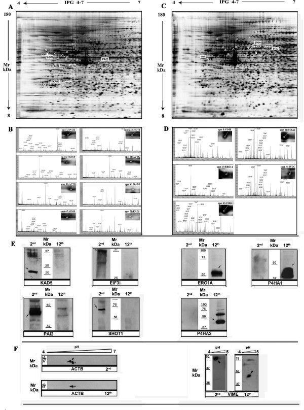
Results: To better understand their self-renewal and potential in vitro expansion capacity, a reference 2D map was constructed as a proteomic data set. 158 unique proteins were identified. More than 30% of these proteins belong to cytoskeleton compartment. We also found that several proteins including Shootin1, Adenylate kinase 5 isoenzyme and Plasminogen activator-inhibitor 2 are no longer expressed after the 2nd passage of in vitro replication. This indicates that the proliferative potency of these cells is reduced after the initial stage of in vitro growing. At the end of cellular culturing, new synthesized proteins, including, ERO1-like protein alpha, Aspartyl-tRNA synthetase and Prolyl-4-hydroxylase were identified. It is suggested that these new synthesized proteins are involved in the impairment of cellular surviving during replication and differentiation time.
Conclusions: Our work represents an essential step towards gaining knowledge of the molecular properties of WJCs so as to better understand their possible use in the field of cell therapy and regenerative medicine.
Figures

Similar articles
-
Matrix cells from Wharton's jelly form neurons and glia.Stem Cells. 2003;21(1):50-60. doi: 10.1634/stemcells.21-1-50. Stem Cells. 2003. PMID: 12529551
-
Human umbilical cord Wharton's Jelly-derived mesenchymal stem cells differentiation into nerve-like cells.Chin Med J (Engl). 2005 Dec 5;118(23):1987-93. Chin Med J (Engl). 2005. PMID: 16336835
-
Human Wharton's Jelly-Cellular Specificity, Stemness Potency, Animal Models, and Current Application in Human Clinical Trials.J Clin Med. 2020 Apr 12;9(4):1102. doi: 10.3390/jcm9041102. J Clin Med. 2020. PMID: 32290584 Free PMC article. Review.
-
In vitro differentiation process of human Wharton's jelly mesenchymal stem cells to male germ cells in the presence of gonadal and non-gonadal conditioned media with retinoic acid.In Vitro Cell Dev Biol Anim. 2015 Nov;51(10):1093-101. doi: 10.1007/s11626-015-9929-4. Epub 2015 Oct 1. In Vitro Cell Dev Biol Anim. 2015. PMID: 26427713
-
New emerging potentials for human Wharton's jelly mesenchymal stem cells: immunological features and hepatocyte-like differentiative capacity.Stem Cells Dev. 2010 Apr;19(4):423-38. doi: 10.1089/scd.2009.0299. Stem Cells Dev. 2010. PMID: 19958166 Review.
Cited by
-
Umbilical Cord Tissue-Derived Cells as Therapeutic Agents.Stem Cells Int. 2015;2015:150609. doi: 10.1155/2015/150609. Epub 2015 Jul 12. Stem Cells Int. 2015. PMID: 26246808 Free PMC article. Review.
-
Efficient improvement of the proliferation, differentiation, and anti-arthritic capacity of mesenchymal stem cells by simply culturing on the immobilized FGF2 derived peptide, 44-ERGVVSIKGV-53.J Adv Res. 2024 Aug;62:119-141. doi: 10.1016/j.jare.2023.09.041. Epub 2023 Sep 28. J Adv Res. 2024. PMID: 37777063 Free PMC article.
-
The role of the adenylate kinase 5 gene in various diseases and cancer.J Clin Transl Sci. 2024 Aug 19;8(1):e96. doi: 10.1017/cts.2024.536. eCollection 2024. J Clin Transl Sci. 2024. PMID: 39655021 Free PMC article. Review.
-
Proteomic Characterization of Changes in Mouse Brain Cortex Protein Expression at Different Post-Mortem Intervals: A Preliminary Study for Forensic Biomarker Identification.Int J Mol Sci. 2024 Aug 10;25(16):8736. doi: 10.3390/ijms25168736. Int J Mol Sci. 2024. PMID: 39201424 Free PMC article.
-
Stem Cell Banking for Regenerative and Personalized Medicine.Biomedicines. 2014 Feb 26;2(1):50-79. doi: 10.3390/biomedicines2010050. Biomedicines. 2014. PMID: 28548060 Free PMC article. Review.
References
-
- Friedenstein AJ, Chailakhjan RK, Lalykina KS. The development of fibroblast colonies in monolayer cultures of guinea-pig bone marrow and spleen cells. Cell Tissue Kinet. 1970;3:393–403. - PubMed
-
- Sacchetti B, Funari A, Michienzi S, Di Cesare S, Piersanti S, Saggio I, Tagliafico E, Ferrari S, Robey PG, Riminucci M, Bianco P. Self-renewing osteoprogenitors in bone marrow sinusoids can organize a hematopoietic microenvironment. Cell. 2007;131:324–336. doi: 10.1016/j.cell.2007.08.025. - DOI - PubMed
LinkOut - more resources
Full Text Sources
