

Transport and biodistribution of dendrimers across human fetal membranes: implications for intravaginal administration of dendrimer-drug conjugates
- PMID: 20346497
- PMCID: PMC2881551
- DOI: 10.1016/j.biomaterials.2010.02.075
Transport and biodistribution of dendrimers across human fetal membranes: implications for intravaginal administration of dendrimer-drug conjugates
Abstract
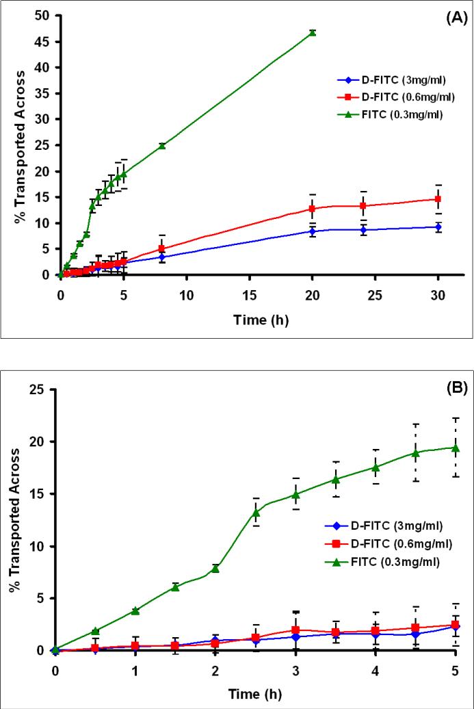
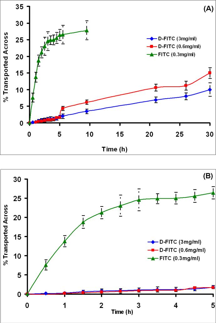
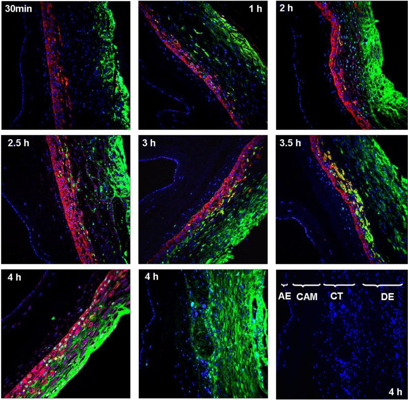
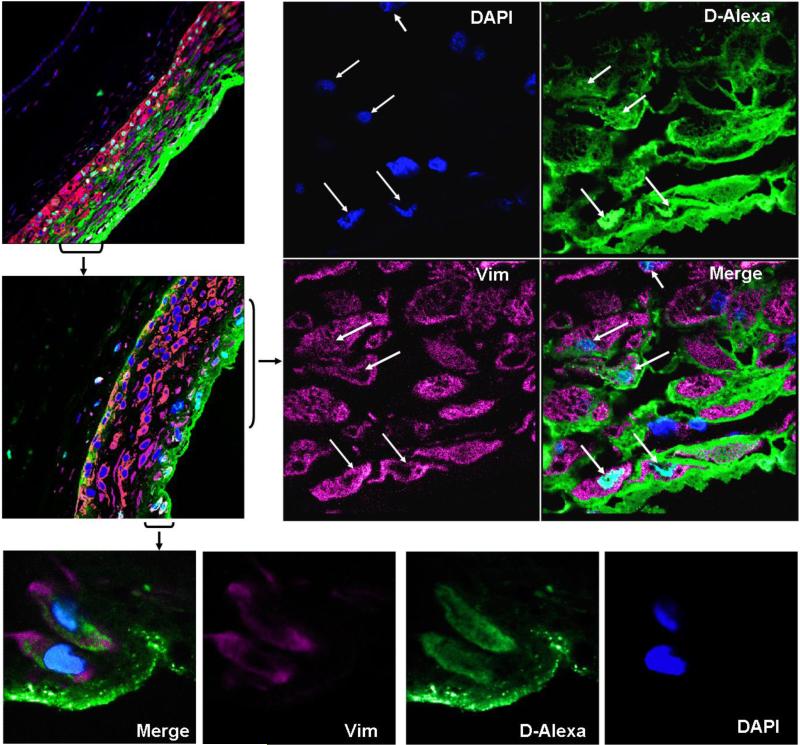
Dendrimers are emerging as promising topical antimicrobial agents, and as targeted nanoscale drug delivery vehicles. Topical intravaginal antimicrobial agents are prescribed to treat the ascending genital infections in pregnant women. The fetal membranes separate the extra-amniotic space and fetus. The purpose of the study is to determine if the dendrimers can be selectively used for local intravaginal application to pregnant women without crossing the membranes into the fetus. In the present study, the transport and permeability of PAMAM (poly (amidoamine)) dendrimers, across human fetal membrane (using a side by side diffusion chamber), and its biodistribution (using immunofluorescence) are evaluated ex-vivo. Transport across human fetal membranes (from the maternal side) was evaluated using Fluorescein (FITC), an established transplacental marker (positive control, size approximately 400 Da) and fluorophore-tagged G(4)-PAMAM dendrimers (approximately 16 kDa). The fluorophore-tagged G(4)-PAMAM dendrimers were synthesized and characterized using (1)H NMR, MALDI TOF MS and HPLC analysis. Transfer was measured across the intact fetal membrane (chorioamnion), and the separated chorion and amnion layers. Over a 5 h period, the dendrimer transport across all the three membranes was less than <3%, whereas the transport of FITC was relatively fast with as much as 49% transport across the amnion. The permeability of FITC (7.9 x 10(-7) cm(2)/s) through the chorioamnion was 7-fold higher than that of the dendrimer (5.8 x 10(-8) cm(2)/s). The biodistribution showed that the dendrimers were largely present in interstitial spaces in the decidual stromal cells and the chorionic trophoblast cells (in 2.5-4 h) and surprisingly, to a smaller extent internalized in nuclei of trophoblast cells and nuclei and cytoplasm of stromal cells. Passive diffusion and paracellular transport appear to be the major route for dendrimer transport. The overall findings further suggest that entry of drugs conjugated to dendrimers would be restricted across the human fetal membranes when administered topically by intravaginal route, suggesting new ways of selectively delivering therapeutics to the mother without affecting the fetus.
(c) 2010 Elsevier Ltd. All rights reserved.
Figures


Similar articles
-
Transfer of PAMAM dendrimers across human placenta: prospects of its use as drug carrier during pregnancy.J Control Release. 2011 Mar 30;150(3):326-38. doi: 10.1016/j.jconrel.2010.11.023. Epub 2010 Dec 1. J Control Release. 2011. PMID: 21129423 Free PMC article.
-
Comparative study of the human amnion, chorion and chorioamnion permeability to monovalent cations.Eur J Obstet Gynecol Reprod Biol. 1983 Sep;16(1):1-7. doi: 10.1016/0028-2243(83)90213-7. Eur J Obstet Gynecol Reprod Biol. 1983. PMID: 6313442
-
Fetal uptake of intra-amniotically delivered dendrimers in a mouse model of intrauterine inflammation and preterm birth.Nanomedicine. 2014 Aug;10(6):1343-51. doi: 10.1016/j.nano.2014.03.008. Epub 2014 Mar 19. Nanomedicine. 2014. PMID: 24657482
-
Expand classical drug administration ways by emerging routes using dendrimer drug delivery systems: a concise overview.Adv Drug Deliv Rev. 2013 Oct;65(10):1316-30. doi: 10.1016/j.addr.2013.01.001. Epub 2013 Feb 13. Adv Drug Deliv Rev. 2013. PMID: 23415951 Review.
-
Poly(amido amine) dendrimers in oral delivery.Tissue Barriers. 2016 Apr 6;4(2):e1173773. doi: 10.1080/21688370.2016.1173773. eCollection 2016 Apr-Jun. Tissue Barriers. 2016. PMID: 27358755 Free PMC article. Review.
Cited by
-
Gestational nanomaterial exposures: microvascular implications during pregnancy, fetal development and adulthood.J Physiol. 2016 Apr 15;594(8):2161-73. doi: 10.1113/JP270581. Epub 2015 Oct 28. J Physiol. 2016. PMID: 26332609 Free PMC article.
-
Rational chemical design of the next generation of molecular imaging probes based on physics and biology: mixing modalities, colors and signals.Chem Soc Rev. 2011 Sep;40(9):4626-48. doi: 10.1039/c1cs15077d. Epub 2011 May 23. Chem Soc Rev. 2011. PMID: 21607237 Free PMC article. Review.
-
Inhibition of bacterial growth and intramniotic infection in a guinea pig model of chorioamnionitis using PAMAM dendrimers.Int J Pharm. 2010 Aug 16;395(1-2):298-308. doi: 10.1016/j.ijpharm.2010.05.030. Epub 2010 May 24. Int J Pharm. 2010. PMID: 20580797 Free PMC article.
-
Structure-skin permeability relationship of dendrimers.Pharm Res. 2011 Sep;28(9):2246-60. doi: 10.1007/s11095-011-0455-0. Epub 2011 Jun 2. Pharm Res. 2011. PMID: 21633876
-
Designing dendrimers for drug delivery and imaging: pharmacokinetic considerations.Pharm Res. 2011 Jul;28(7):1500-19. doi: 10.1007/s11095-010-0339-8. Epub 2010 Dec 23. Pharm Res. 2011. PMID: 21181549 Review.
References
-
- Audus KL. Controlling drug delivery across the placenta. Eur J Pharm Sci. 1999;8(3):161–165. - PubMed
-
- Sastry BV. Techniques to study human placental transport. Adv Drug Deliv Rev. 1999;38(1):17–39. - PubMed
-
- Chaim W, Mazor M, Leiberman JR. The relationship between bacterial vaginosis and preterm birth. A review. Arch Gynecol Obstet. 1997;259:51–58. - PubMed
Publication types
MeSH terms
Substances
Grants and funding
LinkOut - more resources
Full Text Sources
Other Literature Sources
