. 2010 Jun;31(18):4880-8.
doi: 10.1016/j.biomaterials.2010.02.055. Epub 2010 Mar 25.

A defined long-term in vitro tissue engineered model of neuromuscular junctions

Affiliations

A defined long-term in vitro tissue engineered model of neuromuscular junctions

Mainak Das et al. Biomaterials. 2010 Jun.

Abstract

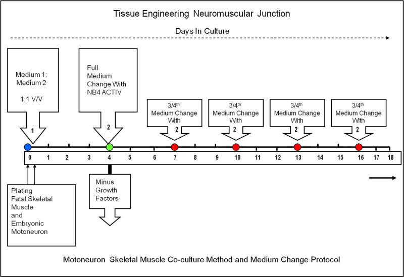
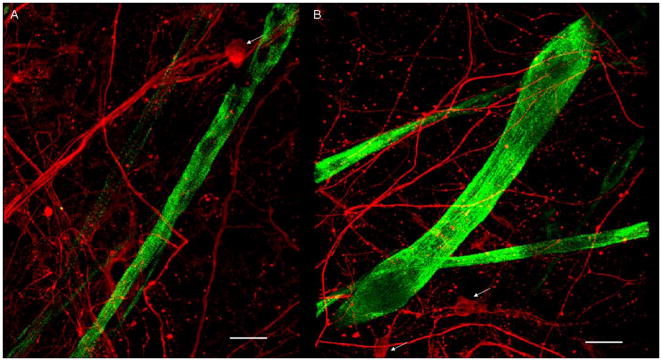
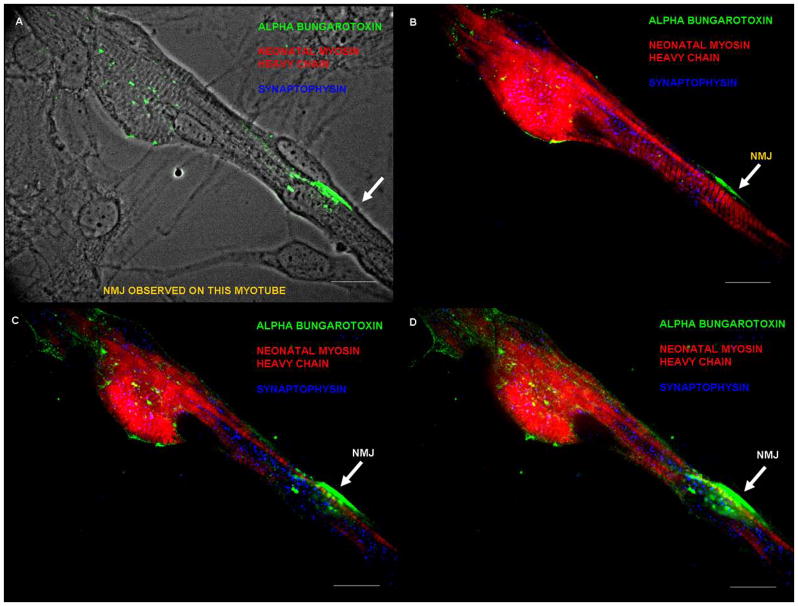
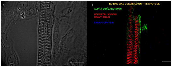
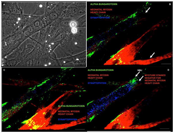
Neuromuscular junction (NMJ) formation, occurring between motoneurons and skeletal muscle, is a complex multistep process involving a variety of signaling molecules and pathways. In vitro motoneuron-muscle co-cultures are powerful tools to study the role of different growth factors, hormones and cellular structures involved in NMJ formation. In this study, a serum-free culture system utilizing defined temporal growth factor application and a non-biological substrate resulted in the formation of robust NMJs. The system resulted in long-term survival of the co-culture and selective expression of neonatal myosin heavy chain, a marker of myotube maturation. NMJ formation was verified by colocalization of dense clusters of acetylcholine receptors visualized using alpha-bungarotoxin and synaptophysin containing vesicles present in motoneuron axonal terminals. This model will find applications in basic NMJ research and tissue engineering applications such as bio-hybrid device development for limb prosthesis and regenerative medicine as well as for high-throughput drug and toxin screening applications.


Figures

Figure 1
Figure 1
Protocol for long-term NMJ formation in a motoneuron and skeletal muscle co-culture.
Figure 2
Figure 2. Phase contrast micrographs of the motoneuron and skeletal muscle co-culture between days 12–15
(A–D) Red arrows indicated the distinct morphology of the motoneuron and its processes. Green arrows indicate the myotubes. Scale bar = 25 μm.
Figure 3
Figure 3. Phase contrast pictures of the co-cultures between days 25–30
(A, B) The myotubes exhibited characteristic striations. (C, D) Myotubes with striations and myotubes without striations. Red arrows indicate the motoneuron cell body and the processes. The green arrows indicate the myotubes. The scale bar for A, B = 40 μm. The scale bar for C, D = 25 μm.
Figure 4
Figure 4. Immunocytochemistry of co-cultures at day 25
(A–B) NF-150 (red) indicates the large motoneurons and their processes (white arrows). The striated myotubes (green) stained for nMHC (N3.36). Scale bar = 50 μm.
Figure 5
Figure 5. Neuromuscular junction (NMJ) formation between day 30–40
(A) Phase picture of the myotube indicating the alpha-bungarotoxin staining in green, (B) Triple stain, showing the close proximity of alpha-bungarotoxin (green) and synaptophysin (blue) indicating synapse formation at a specific confocal plane and myotube striations are indicated in red (nMHC), (C–D) NMJ observed at two different planes using confocal microscopy. A much more dense clustering of synaptophysin and alpha-bungarotoxin was observed in these planes.
Figure 6
Figure 6. Striated myotube development in the absence of NMJ formation
(A, B) No NMJs were observed on this striated myotube. (A) A phase picture of the myotube, (B) Immunostained picture of the same myotube with alpha-bungarotoxin, N3.36 and synaptophysin. Scale bar = 50 μm.
Figure 7
Figure 7. NMJ formation on a N3.36 (−) myotube
(A) Phase picture showing the different morphologies of myotubes in the co-culture, (B–D) NMJ formation was observed on a myotube that was negative for N3.36. Culture stained with alpha-bungarotoxin, N3.36 and synaptophysin. Possibly the myotube on which NMJ was formed was immature and had not yet expressed the neonatal myosin heavy chain marker (N3.36).

Similar articles

Cited by

References

    1. Witzemann V. Development of the neuromuscular junction. Cell Tissue Res. 2006;326(2):263–71. - PubMed
    1. Chow I, Poo MM. Release of acetylcholine from embryonic neurons upon contact with muscle cell. J Neurosci. 1985;5(4):1076–82. - PMC - PubMed
    1. Colomar A, Robitaille R. Glial modulation of synaptic transmission at the neuromuscular junction. Glia. 2004;47(3):284–9. - PubMed
    1. English AW. Cytokines, growth factors and sprouting at the neuromuscular junction. J Neurocytol. 2003;32(5):943–60. - PubMed
    1. Ravenscroft MS, Bateman KE, Shaffer KM, Schessler HM, Jung DR, Schneider TW, et al. Developmental neurobiology implications from fabrication and analysis of hippocampal neuronal networks on patterned silane- modified surfaces. J Am Chem Soc. 1998;120(47):12169–77.

Publication types

Substances