Review
. 2010 Aug;35(8):434-41.
doi: 10.1016/j.tibs.2010.03.001. Epub 2010 Mar 26.

The molecular regulation of programmed necrotic cell injury

Affiliations
Review

The molecular regulation of programmed necrotic cell injury

David Moquin et al. Trends Biochem Sci. 2010 Aug.

Abstract

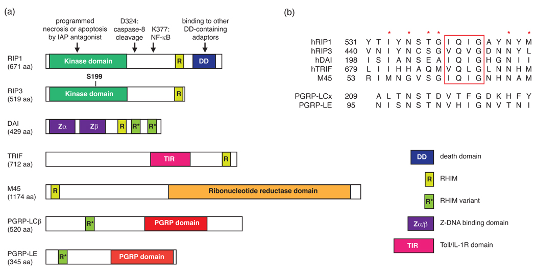
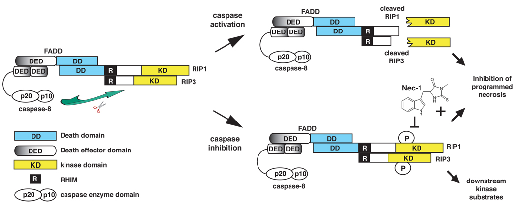
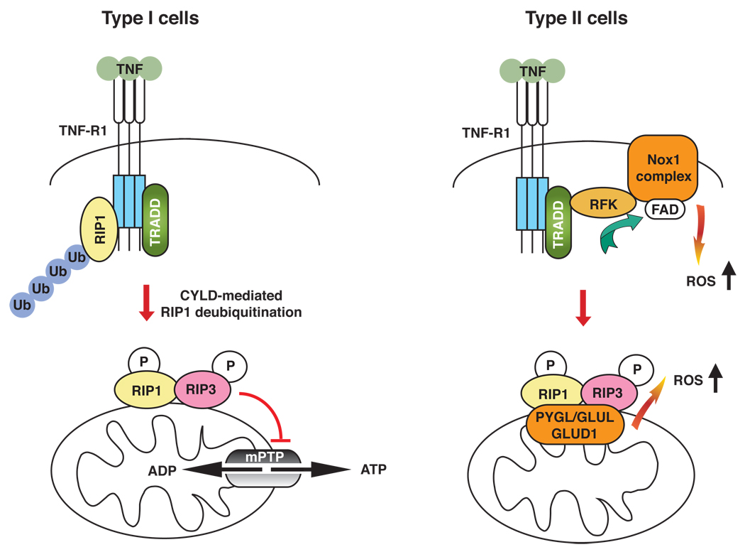
Proper regulation of cell death is essential for metazoan development and functions. Unlike apoptosis, necrosis is a more inflammatory form of cell death that might contribute to antiviral immunity. Indeed, necrotic cell injury is distinguished from apoptosis by extensive organelle and cell swelling and plasma membrane rupture. Recent evidence indicates that an elaborate biochemical network emanating from receptors in the TNF superfamily can induce apoptosis as well as necrotic cell death. The induction of necrosis by TNF-like cytokines requires biochemical components that are distinct from those involved in apoptosis. Specifically, serine/threonine protein kinases in the receptor interacting protein (RIP) family are required for "programmed" necrotic cell injury. In this review, we discuss the molecular crosstalk between apoptosis and programmed necrosis, with a special emphasis on how caspases, protein ubiquitylation and phosphorylation regulate the induction of necrotic cell injury.


Figures

Figure 1
Figure 1. The emerging family of RHIM-containing proteins
(a) Schematic diagram of human, viral and Drosophila adaptor proteins containing RHIMs. The arrows indicate the amino acid residues and domains important for RIP1 function. S199 is a reported necrosis-specific phosphorylation target site on RIP3 [28]. (b) Sequence alignment of the RHIM or RHIM-like domains. The red rectangle denotes the highly conserved (I/V)Q(I/V/L)G tetrapeptide found in the RHIM. The red asterisks represent other highly conserved residues within the RHIM domain. The numbers represent the amino-terminal boundary of the listed sequences.
Figure 2
Figure 2. Caspase 8-mediated cleavage of RIP1 and RIP3 inhibits programmed necrosis
The assembly of a cytoplasmic RIP1–RIP3 complex via the RHIM is critical for programmed necrosis. This complex is further stabilized by phosphorylation of RIP1 and RIP3. FADD and caspase 8 inhibit TNF-induced programmed necrosis by cleaving RIP1 (at D324) and RIP3 (at D333) within this complex. Cleavage results in the release of amino-terminal fragments containing the kinase domains, thereby preventing phosphorylation of RIP1, RIP3 and other possible downstream substrates. Necrostatin-1 (Nec-1), which inhibits RIP1 kinase activity, blocks programmed necrosis by preventing the stable association of the RIP1–RIP3 complex.
Box 2, Figure 1
Box 2, Figure 1. ROS-dependent and independent programmed necrosis in different cell types
Assembly of the RIP1–RIP3 (pink–yellow) complex involves deubiquitylation of RIP1, possibly by CYLD. The assembled RIP1–RIP3 complex might act on the mitochondria to trigger downstream effector functions. In Type I cells such as the U937 cell line, Jurkat T-cells, and the HT-29 cell line, ROS scavengers such as butylated hydroxyanisole (BHA) do not rescue programmed necrosis. In these cells, programmed necrosis might be mediated by loss of function of the mPTP and cellular ATP. By contrast, in Type II cells such as the L929 cell line or MEFs, programmed necrosis is ROS-dependent as BHA rescues the cell death. Evidence indicates that ROS can be produced at the RFK–NOX1–FAD plasma membrane complex or within the mitochondria through RIP3-mediated interaction with mitochondrial metabolic enzymes (PYGL, GLUL, GLUD1; orange). It is unclear if the two sources of ROS might synergize with each other to facilitate programmed necrosis. Ubiquitin (Ub): blue.

Similar articles

Cited by

References

    1. Degterev A, et al. Identification of RIP1 kinase as a specific cellular target of necrostatins. Nat Chem Biol. 2008;4:313–321. - PMC - PubMed
    1. Degterev A, et al. Chemical inhibitor of nonapoptotic cell death with therapeutic potential for ischemic brain injury. Nat Chem Biol. 2005;1:112–119. - PubMed
    1. Tu HC, et al. The p53-cathepsin axis cooperates with ROS to activate programmed necrotic death upon DNA damage. Proceedings of the National Academy of Sciences of the United States of America. 2009;106:1093–1098. - PMC - PubMed
    1. Papa S, et al. Linking JNK signaling to NF-kappaB: a key to survival. Journal of cell science. 2004;117:5197–5208. - PubMed
    1. Carswell EA, et al. An endotoxin-induced serum factor that causes necrosis of tumors. Proceedings of the National Academy of Sciences of the United States of America. 1975;72:3666–3670. - PMC - PubMed

Publication types

Substances

LinkOut - more resources