. 2010 Apr 23;38(2):165-78.
doi: 10.1016/j.molcel.2010.03.002. Epub 2010 Mar 25.

A functional link between the histone demethylase PHF8 and the transcription factor ZNF711 in X-linked mental retardation

Affiliations

A functional link between the histone demethylase PHF8 and the transcription factor ZNF711 in X-linked mental retardation

Daniela Kleine-Kohlbrecher et al. Mol Cell. .

Abstract

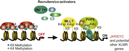
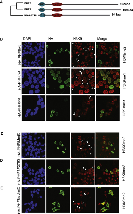
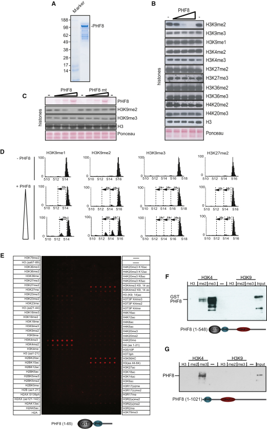
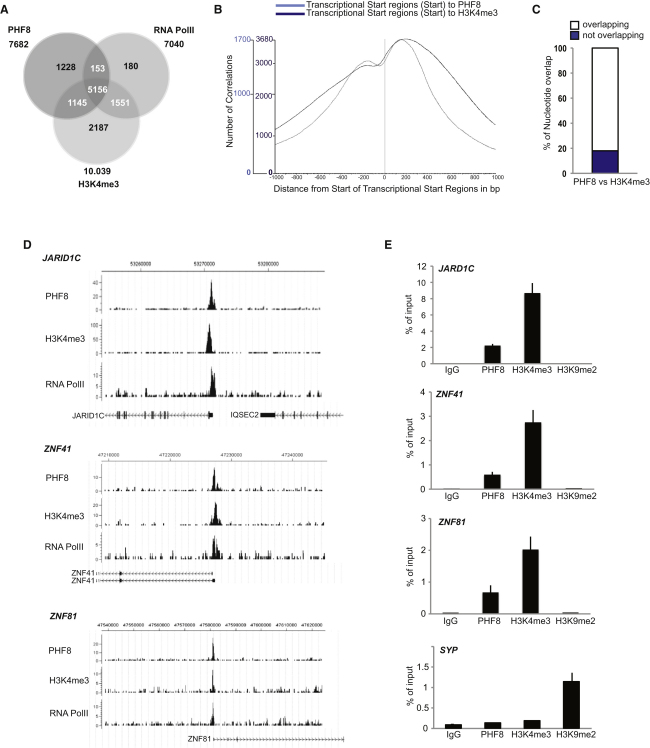
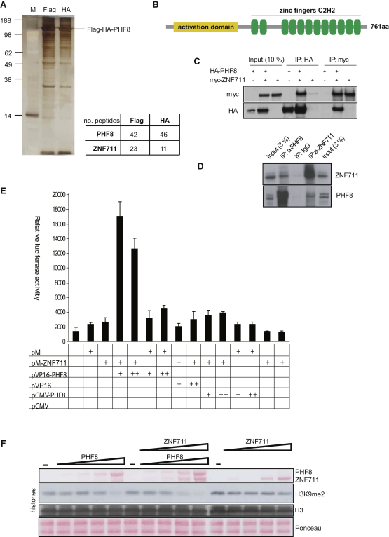
X-linked mental retardation (XLMR) is an inherited disorder that mostly affects males and is caused by mutations in genes located on the X chromosome. Here, we show that the XLMR protein PHF8 and a C. elegans homolog F29B9.2 catalyze demethylation of di- and monomethylated lysine 9 of histone H3 (H3K9me2/me1). The PHD domain of PHF8 binds to H3K4me3 and colocalizes with H3K4me3 at transcription initiation sites. Furthermore, PHF8 interacts with another XMLR protein, ZNF711, which binds to a subset of PHF8 target genes, including the XLMR gene JARID1C. Of interest, the C. elegans PHF8 homolog is highly expressed in neurons, and mutant animals show impaired locomotion. Taken together, our results functionally link the XLMR gene PHF8 to two other XLMR genes, ZNF711 and JARID1C, indicating that MR genes may be functionally linked in pathways, causing the complex phenotypes observed in patients developing MR.


Figures

None
Graphical abstract
Figure 1
Figure 1
PHF8 Demethylates H3K9me2 In Vivo (A) Phylogenetic analysis and schematic representation of the human PHF family. Alignment of the proteins and construction of the phylogenetic tree were conducted using the ClustalW (http://align.genome.jp). Shown are jumonji C domain (JmjC) and plant homeotic domain (PHD). (B–E) (B) 293 cells were transfected with HA-tagged PHF8, (C) a PHF JmjC mutant, (D) a naturally occurring XLMR mutant with mutation in the JmjC domain (F279S), and (E) a C-terminal deletion (K177X) found in XLMR patients. The transfected cells were fixed, costained for the indicated histone modification and for the expression of the proteins (anti-HA), and analyzed by confocal microscopy. White arrows indicate cells expressing the tested protein. The cells were counterstained with DAPI to visualize cell nuclei.
Figure 2
Figure 2
PHF8 Demethylates H3K9me2/me1 In Vitro and Binds to H3K4me3 (A) SDS-PAGE analysis of purified His-tagged recombinant PHF8 expressed in insect cells. Marker is a molecular weight standard. Samples were subjected to SDS-PAGE and stained using Coomassie blue. The arrow indicates the position of recombinant PHF8 (120 kDa). (B) Demethylation assays of histones incubated with 1.5, 4.5, and 12.5 μg of recombinant PHF8; bottom panels show Ponceau stain of the histones present in the assays. The middle panels show the reactions probed with the indicated antibodies and assayed by immunoblotting. (C) Demethylation assays of histones incubated with 1.5, 4.5, and 12.5 μg of recombinant PHF8 or a PHF8 JmjC mutant (H247R); bottom panels show Ponceau stain of the histones present in the assays. The middle panels show the reactions probed with the indicated antibodies and assayed by immunoblotting. (D) H3K9me3/me2/me1 and H3K27me2 peptides (2.5 μg) were incubated with or without recombinant PHF8 (0.5 or 1.5 μg) and analyzed by mass spectrometry. A shift in mass equivalent to one methyl group is indicated as “Me.” (E) Peptide array spotted with six replicates of modified peptides covering the large parts of histone H2A, H2B, H3, H2AX, and H4 modified by different degrees and combinations of methylation, acetylation, and phosphorylation. The modifications are listed in the panel. The array was incubated with a GST fusion protein harboring the PHD finger of PHF8, and the detection of bound protein was performed as described in the Supplemental Experimental Procedures. (F) In vitro binding experiment. A purified GST-PHF8 fusion protein (amino acids 1–548) containing the PHD and JmjC domain was incubated with biotinylated histone H3 peptides methylated at either the K4 or K9 position. After precipitation with Streptavidin-agarose, bound PHF8 was detected by SDS-PAGE and immunoblotting. (G) In vitro binding experiments. A lysate from HeLa cells was incubated with biotinylated histone H3 peptides methylated at either the K4 or K9 position. After precipitation with Streptavidin-agarose, bound PHF8 was detected by SDS-PAGE and immunoblotting.
Figure 3
Figure 3
PHF8 Binds Target Genes in the H3K4me3-Positive Region Surrounding the TSS (A) Venn diagram showing the overlap and number of genes positive for PHF8, H3K4me3, and RNA pol II in human neuroblastoma SH-SY5Y cells. (B) Distribution of the distance between PHF8 (light blue) and H3K4me2 (dark blue) peaks and the transcription start sites (TSS). (C) PHF8 nucleotides that are also bound by H3K4me3. (D) Binding profiles of the XLMR genes JARID1C, ZNF41, and ZNF81 for PHF8, H3K4me3, and RNA pol II obtained from ChIP-seq analysis; the chromosomal locations are indicated in the top of the panel according to hg18. The Y axes indicate the number of sequenced reads. (E) qChIP analysis of the XMLR genes JARID1C, ZNF41, and ZNF81 in SH-SY5Y cells using antibodies against PHF8, H3K4me3, H3K9me2, and IgG control. The enrichment of the ChIP assays is shown as percentage of input. qChIP analysis of the gene coding for synaptophysin (SYP) using antibodies against PHF8, H3K4me3, H3K9me2, and IgG (control) was used as positive control for H3K9me2. Error bars represent SD; n = 3.
Figure 4
Figure 4
PHF8 Interacts with ZNF711 (A) Flag-HA tandem purification of PHF8 complexes. Silver-stained SDS-PAGE analysis of Flag- and Flag-HA-purified complex from 293 cells. M, molecular weight marker; F, Flag-eluate; H, Flag-eluate further purified by HA-affinity chromatography. The eluted material was analyzed in bulk by mass spectrometry. The numbers of peptides identified for PHF8 and ZNF11 for single-step Flag purification or double Flag-HA purification are shown. (B) Schematic representation of ZNF711. Shown are the putative activation domain and ZNF, Cys-His zinc finger domain. (C) Coimmunoprecipitation of PHF8 and ZNF711. Phoenix cells were transfected or cotransfected with the expression vectors pCMV-HA-PHF8 and pCMV-Myc-ZNF711 and were immunoprecipitated using antibodies against the HA-tag or Myc-tag as indicated. The precipitates were analyzed by SDS-PAGE followed by western blotting using antibodies against the Myc-tag or HA-tag. (D) Coimmunoprecipitation of endogenous PHF8 and ZNF711. HEK293 cells were lysed and immunoprecipitated with the indicated antibodies against ZNF711 and PHF8. The precipitates were analyzed by SDS-PAGE followed by western blotting using antibodies against ZNF711 and PHF8. (E) Interaction of PHF8 and ZNF711 in a mammalian two-hybrid assay. U2OS cells were transfected with a pGAL-Luc reporter plasmid containing the luciferase gene driven by the adenovirus E1B minimal promoter (TATA) fused to five upstream GAL4-binding sites or cotransfected with the expression constructs indicated in the bottom of the panel. For correction of transfection efficiency, pCMV-lacZ was included in all assays, and luciferase activity was normalized to β-galactosidase activity. All experiments were performed in triplicate and reproduced at least three times. Error bars represent SD; n = 3. (F) Demethylation assays of histones incubated with 1.5, 4.5, and 12.5 μg of recombinant PHF8 alone and/or in the presence of 1, 3, and 9 ug of recombinant ZNF711; bottom panels show Ponceau stain of the histones present in the assays. The middle panels show the reactions probed with the indicated antibodies and assayed by immunoblotting.
Figure 5
Figure 5
PHF8 Colocalizes with ZNF711 on Target Genes (A) Venn diagram showing the overlap and number of genes positive for ZNF711, PHF8, and H3K4me3 in human neuroblastoma SH-SY5Y cells. (B) Binding profiles of the ZNF711 target genes JARID1C, C2ORF34, and PCBP2 for ZNF711 and PHF8 obtained from ChIP-seq analysis; the chromosomal locations are indicated in the top of the panel according to hg18. The Y axes indicate the number of sequenced reads. (C) qChIP analysis of ZNF711 target genes JARID1C, C2ORF34, and PCBP2 in SH-SY5Y cells using antibodies against ZNF711, PHF8, H3K4me3, H3K9me2, and IgG control. The enrichment of the ChIP assays is shown as percentage bound of input. (D) qChIP analysis of ZNF711 target genes JARID1C and C2ORF34 in HEK293 cells after shRNA-mediated ZNF711 knockdown using antibodies against ZNF711, PHF8, H3K4me3, and IgG control. (E) Expression analysis of ZNF711-PHF8 target genes after shRNA-mediated ZNF711 and PHF8 knockdown in HEK293 cells using real-time RT-qPCR. Error bars represent SD; n = 3.
Figure 6
Figure 6
The C. elegans PHF8 Homolog F29B9.2 Is an H3K9me2 and H3K27me2 Demethylase Involved in Coordinated Movement (A) (Top) Schematic representation of the C. elegans F29B9.2 protein. PHD, plant homeodomain finger; JmjC, jumonji C domain. (Bottom) Genomic organization of the F29B9.2 gene. Exons are indicated by black boxes. Black H-shaped line indicates the position of the tm3713 deletion. Black line indicates the position of the fragment used for RNA interference. (B) SDS-PAGE analysis of purified His-tagged recombinant F29B9.2 expressed in insect cells. Marker is molecular weight standard. Samples were subjected to SDS-PAGE and stained using Coomassie blue. The arrow indicates the position of recombinant F29B9.2. Demethylation assays of histones incubated with 1.5 and 4.5 μg of recombinant F29B9.2; bottom panels show Ponceau stain of the histones present in the assays. The middle panels show the reactions probed with the indicated antibodies and assayed by immunoblotting. (C) Analysis of histone modifications in wild-type animals (N2), tm3713 mutants, and RNAi (F29B9.2)-treated animals. (Left) Protein lysates from synchronized N2 and tm3713 young adults were probed with the indicated antibodies. (Middle) Protein lysates from synchronized F1 eri-1 adult worms treated with control or F29B9.2(RNAi) were probed with antibodies as indicated. (Right) The relative expression level of F29B9.2 mRNA in control and RNAi-treated animals, quantified by real-time RT-PCR. Error bars represent SD; n = 3. (D) Epifluorescent image of an adult hermaphrodite carrying the F29B9.2::GFP transgene. VC, ventral nerve cord; AG, anterior ganglia; PG, posterior ganglia; E, embryos. In (D) and (F), anterior is in the left; ventral, down. Scale bar represents 100 μm. (E) Abnormal movement on bacterial-seeded plates of tm3713 mutant animals, rescued in tm3713 carrying the F29B9.2::GFP transgene (tm3713R). The graphics show the measure of the wavelength and amplitude of N2, tm3713, and tm3713R. Values were normalized to the N2. Error bars in (E) and (F) represent standard deviation of the mean. Asterisks in the graphics indicate results different at p < 0.01 (Student's t test). The variations on amplitude are not significant (p > 0.05). (F) tm3713 mutant animals expressing F29B9.2 under neuronal (rab-3) and muscle (myo-3) promoters. Expression of F29B9.2 in neurons restores wild-type locomotion. The graphic shows the measure of the wavelength of N2 and transgenic animals normalized to the values of the N2. (G) PHF8, ZNF711, and JARID1C are linked in a pathway required for suppressing mental retardation.

Comment in

  • Demethylases go mental.
    Dawson MA, Bannister AJ. Dawson MA, et al. Mol Cell. 2010 Apr 23;38(2):155-7. doi: 10.1016/j.molcel.2010.04.002. Mol Cell. 2010. PMID: 20417593 No abstract available.

Similar articles

Cited by

References

    1. Abidi F.E., Miano M.G., Murray J.C., Schwartz C.E. A novel mutation in the PHF8 gene is associated with X-linked mental retardation with cleft lip/cleft palate. Clin. Genet. 2007;72:19–22. - PMC - PubMed
    1. American Psychiatric Association . Fourth Edition. The American Psychiatric Association; Washington, DC: 1994. Diagnostic and statistical manual of mental disorders.
    1. Barski A., Cuddapah S., Cui K., Roh T.Y., Schones D.E., Wang Z., Wei G., Chepelev I., Zhao K. High-resolution profiling of histone methylations in the human genome. Cell. 2007;129:823–837. - PubMed
    1. Bracken A.P., Dietrich N., Pasini D., Hansen K.H., Helin K. Genome-wide mapping of Polycomb target genes unravels their roles in cell fate transitions. Genes Dev. 2006;20:1123–1136. - PMC - PubMed
    1. Champagne K.S., Kutateladze T.G. Structural insight into histone recognition by the ING PHD fingers. Curr. Drug Targets. 2009;10:432–441. - PMC - PubMed

Publication types

MeSH terms

Associated data