. 2010 Jul;136(1):130-8.
doi: 10.1016/j.clim.2010.02.014. Epub 2010 Mar 25.

Human plasmacytoid dendritic cell accumulation amplifies their type 1 interferon production

Affiliations

Human plasmacytoid dendritic cell accumulation amplifies their type 1 interferon production

Anne P Liao et al. Clin Immunol. 2010 Jul.

Abstract

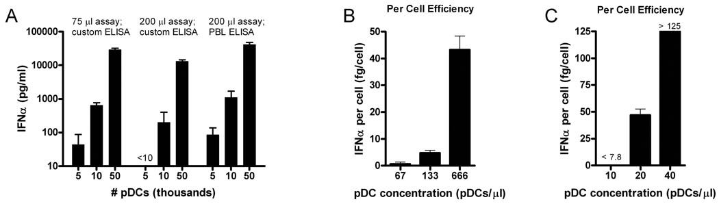
To determine the potential consequences of plasmacytoid dendritic cell (pDC) accumulation in tissue sites observed in several autoimmune diseases, we measured type 1 interferon production from circulating human pDCs as a function of pDC concentration. The effects of interferon-alpha and blockade of the type 1 interferon receptor (IFNAR) on human pDC type 1 interferon and interferon-inducible transcription and protein production were measured. Human pDCs became far more efficient producers of interferon-alpha at concentrations beyond those normally present in blood, through an IFNAR-dependent mechanism. Extracellular interferon-alpha increased pDC production of type 1 interferons. The accumulation of pDCs in diseased tissue sites allows marked non-linear amplification of type 1 interferon production locally. The role of the IFNAR-dependent mechanism of interferon production by human pDCs is greater than previously suggested. IFNAR blockade has potential for diminishing type 1 interferon production by all human cells.


Figures

Figure 1
Figure 1. Non-linearity of pDC interferon-α (IFNα) production demonstrates concentration effects
(A) ODN-stimulated pDC IFNα production increased greater than exponentially, not linearly, with increased cell numbers in 3 different assay formats, in either 75 µl or 200 µl assay volumes or as measured with a custom limited subtype or commercial multisubtype (PBL Biomedical) ELISA. IFNα production plotted with logarithmic scale. (B and C) IFNα production per cell depends on cell density, with marked increases in per cell output with increasing pDC cell concentration. In (B), custom limited subtype ELISA requires large cell numbers for IFNα detection. In (C), custom multisubtype ELISA allows the use of very small numbers of pDCs to study physiologically relevant numbers: 500 pDCs in a 50 µl assay (10 pDCs/µl) approximated the concentration of pDCs in whole blood (5–10 pDCs/µl). Increasing pDC concentration from this value (to 20 and 40 pDC/µl) shows non-linear marked increases in per cell efficiency.
Figure 2
Figure 2. Direct demonstration of IFNα amplification effect on human pDCs
(A) Five different donor pDC collections exposed to TLR9 agonist ODN2216 alone or with 25 pg/ml of IFNα-2a protein. (B) Dose-response effects of IFNα-2a on ODN2216 stimulated pDC production of IFNα. Control pDCs were stimulated with IFNα-2a alone, without ODN2216. 10,000 pDCs, 8 replicates, and custom limited subtype ELISA used for all IFNα measurements.
Figure 3
Figure 3. Kinetics of TLR9 stimulated human pDC type 1 interferon transcription
(A) 24-hour time-scale view of 10 IFNα subtypes, IFNβ, and IFNω transcript levels and their augmentation by the addition of IFNα-2a 25 pg/ml. Intensity = fold change from pre-ODN stimulation levels. (B) Fold-change for 10 subtypes of interferon-α, and interferon-β and –ω transcripts were measured in TLR9 stimulated pDCs in comparison to pDCs prior to stimulation at 1 and 6 hours. Note differing scales, as fold changes have increased substantially at 6 hours.
Figure 4
Figure 4. Effect of type 1 interferon receptor (IFNAR) blockade on IFNα production by human pDCs
(A) Highly sensitive ELISA using low pDC cell numbers shows non-linear increase in IFNα production and that IFNAR blockade (anti-IFNAR2 antibody 5 µg/ml) markedly reduces IFNα production. (B) Lower sensitivity ELISA, 10,000 pDCs per condition. The effects of added IFNα-2a (25 pg/ml) increased IFNα production only in the absence of IFNAR blockade. (p<0.0001 for all comparisons).
Figure 5
Figure 5. Concentrated pDC suspensions produce downstream type 1 interferon-inducible transcripts and proteins, augmented by addition of IFNα
(A) Interferon-inducible transcripts produced by TLR9 stimulated >92% pure pDC suspensions measured by real-time quantitative PCR. Amplification of pDC production of type 1 interferon-inducible transcripts in the presence of small amounts of added IFNα-2a protein (25 pg/ml). 10,000 pDCs in 50 µl volumes used. (B) The interferon-inducible protein Mx1 is present in intracellular fractions and the ubiquitin-like modifier and cytokine protein ISG15 is secreted into cell media at 24 hours. (C) Further downstream effects of type 1 interferon signaling in which multiple intracellular proteins have been conjugated by ISG15 is evident. Unstimulated pDCs (No Tx) do not have or secrete detectable ISG15 or ISG15 conjugates. IFNAR blockade prevents ISG15 conjugation. Transcripts measured by quantitative real-time PCR.
Figure 6
Figure 6. Predicted effects of therapeutic targeting of interferon-alpha (IFNα) compared with targeting the type 1 interferon receptor (IFNAR)
Anti-IFNα neutralization might affect IFNα effects on immune system cells and tissue production of IFN-inducible proteins, but might not affect processes driven by IFNβ or other type 1 interferons. Anti-IFNAR blockade would inhibit (1) tissue production of IFN-inducible proteins regardless of the driving type 1 interferon responsible, (2) the effects of type 1 interferons on activation and maturation of other immune system cells, such as B cells and cytotoxic T cells, and (3) production of all type 1 interferons by pDCs and all other human cells.

References

    1. Pascual V, Farkas L, Banchereau J. Systemic lupus erythematosus: all roads lead to type I interferons. Curr Opin Immunol. 2006;18:676–682. - PubMed
    1. Crow MK. Type I interferon in systemic lupus erythematosus. Curr Top Microbiol Immunol. 2007;316:359–386. - PubMed
    1. Ronnblom L, Alm GV, Eloranta ML. Type I interferon and lupus. Curr Opin Rheumatol. 2009;21:471–477. - PubMed
    1. Greenberg SA. Proposed immunologic models of the inflammatory myopathies and potential therapeutic implications. Neurology. 2007;69:2008–2019. - PubMed
    1. Greenberg SA. Inflammatory myopathies: disease mechanisms. Curr Opin Neurol. 2009;22:516–523. - PubMed

Publication types

MeSH terms

Substances