

The developmental origins of voice processing in the human brain
- PMID: 20346760
- PMCID: PMC2852650
- DOI: 10.1016/j.neuron.2010.03.001
The developmental origins of voice processing in the human brain
Abstract
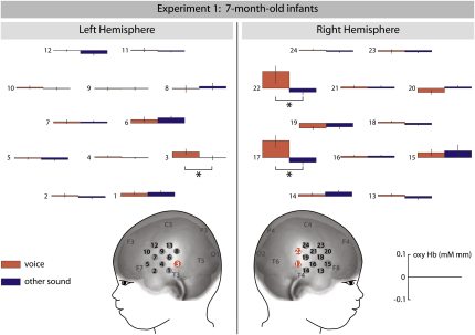
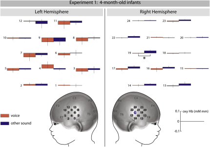
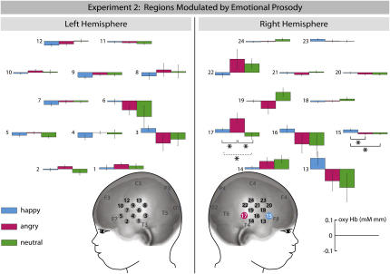
In human adults, voices are processed in specialized brain regions in superior temporal cortices. We examined the development of this cortical organization during infancy by using near-infrared spectroscopy. In experiment 1, 7-month-olds but not 4-month-olds showed increased responses in left and right superior temporal cortex to the human voice when compared to nonvocal sounds, suggesting that voice-sensitive brain systems emerge between 4 and 7 months of age. In experiment 2, 7-month-old infants listened to words spoken with neutral, happy, or angry prosody. Hearing emotional prosody resulted in increased responses in a voice-sensitive region in the right hemisphere. Moreover, a region in right inferior frontal cortex taken to serve evaluative functions in the adult brain showed particular sensitivity to happy prosody. The pattern of findings suggests that temporal regions specialize in processing voices very early in development and that, already in infancy, emotions differentially modulate voice processing in the right hemisphere.
(c) 2010 Elsevier Inc. All rights reserved.
Figures
Comment in
-
Before speech: cerebral voice processing in infants.Neuron. 2010 Mar 25;65(6):733-5. doi: 10.1016/j.neuron.2010.03.018. Neuron. 2010. PMID: 20346748 Review.
Similar articles
-
Voice and emotion processing in the human neonatal brain.J Cogn Neurosci. 2012 Jun;24(6):1411-9. doi: 10.1162/jocn_a_00214. Epub 2012 Feb 23. J Cogn Neurosci. 2012. PMID: 22360593 Clinical Trial.
-
Early specialization for voice and emotion processing in the infant brain.Curr Biol. 2011 Jul 26;21(14):1220-4. doi: 10.1016/j.cub.2011.06.009. Epub 2011 Jun 30. Curr Biol. 2011. PMID: 21723130
-
Differential influences of emotion, task, and novelty on brain regions underlying the processing of speech melody.J Cogn Neurosci. 2009 Jul;21(7):1255-68. doi: 10.1162/jocn.2009.21099. J Cogn Neurosci. 2009. PMID: 18752404
-
Before speech: cerebral voice processing in infants.Neuron. 2010 Mar 25;65(6):733-5. doi: 10.1016/j.neuron.2010.03.018. Neuron. 2010. PMID: 20346748 Review.
-
Processing of emotional vocalizations in bilateral inferior frontal cortex.Neurosci Biobehav Rev. 2013 Dec;37(10 Pt 2):2847-55. doi: 10.1016/j.neubiorev.2013.10.007. Epub 2013 Oct 22. Neurosci Biobehav Rev. 2013. PMID: 24161466 Review.
Cited by
-
Infant pupil diameter changes in response to others' positive and negative emotions.PLoS One. 2011;6(11):e27132. doi: 10.1371/journal.pone.0027132. Epub 2011 Nov 16. PLoS One. 2011. PMID: 22110605 Free PMC article.
-
Isolating N400 as neural marker of vocal anger processing in 6-11-year old children.Dev Cogn Neurosci. 2012 Apr;2(2):268-76. doi: 10.1016/j.dcn.2011.11.007. Epub 2011 Dec 7. Dev Cogn Neurosci. 2012. PMID: 22483076 Free PMC article.
-
Association between neural prosody discrimination and language abilities in toddlers: a functional near-infrared spectroscopy study.BMC Pediatr. 2024 Jul 12;24(1):449. doi: 10.1186/s12887-024-04889-7. BMC Pediatr. 2024. PMID: 38997661 Free PMC article.
-
Emotional voice processing: investigating the role of genetic variation in the serotonin transporter across development.PLoS One. 2013 Jul 8;8(7):e68377. doi: 10.1371/journal.pone.0068377. Print 2013. PLoS One. 2013. PMID: 23861897 Free PMC article.
-
Learning-related brain hemispheric dominance in sleeping songbirds.Sci Rep. 2015 Mar 12;5:9041. doi: 10.1038/srep09041. Sci Rep. 2015. PMID: 25761654 Free PMC article.
References
-
- Aslin R.N., Mehler J. Near-infrared spectroscopy for functional studies of brain activity in human infants: promise, prospects, and challenges. J. Biomed. Opt. 2005;10:11009. - PubMed
-
- Belin P., Zatorre R.J., Lafaille P., Ahad P., Pike B. Voice-selective areas in human auditory cortex. Nature. 2000;403:309–312. - PubMed
-
- Belin P., Fecteau S., Bédard C. Thinking the voice: neural correlates of voice perception. Trends Cogn. Sci. 2004;8:129–135. - PubMed
-
- Blasi A., Fox S., Everdell N., Volein A., Tucker L., Csibra G., Gibson A.P., Hebden J.C., Johnson M.H., Elwell C.E. Investigation of depth dependent changes in cerebral haemodynamics during face perception in infants. Phys. Med. Biol. 2007;52:6849–6864. - PubMed
-
- Borod J.C., Bloom R.L., Brickman A.M., Nakhutina L., Curko E.A. Emotional processing deficits in individuals with unilateral brain damage. Appl. Neuropsychol. 2002;9:23–36. - PubMed
Publication types
MeSH terms
Grants and funding
LinkOut - more resources
Full Text Sources
