

MONICA: a compact, portable dual gamma camera system for mouse whole-body imaging
- PMID: 20346864
- PMCID: PMC2847584
- DOI: 10.1016/j.nucmedbio.2009.12.003
MONICA: a compact, portable dual gamma camera system for mouse whole-body imaging
Abstract
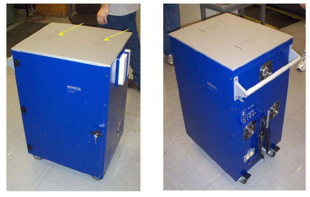
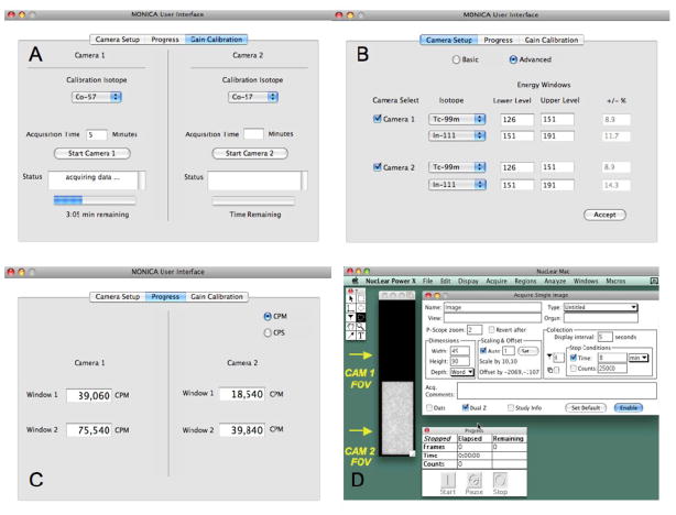
Introduction: We describe a compact, portable dual-gamma camera system (named "MONICA" for MObile Nuclear Imaging CAmeras) for visualizing and analyzing the whole-body biodistribution of putative diagnostic and therapeutic single photon emitting radiotracers in animals the size of mice.
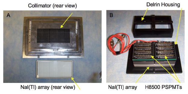
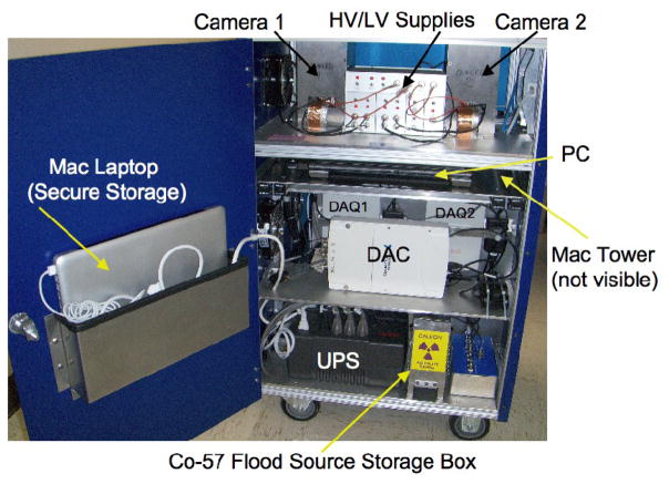
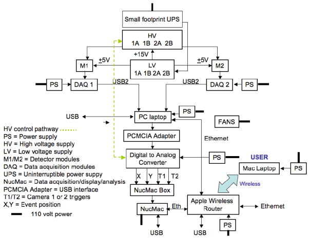
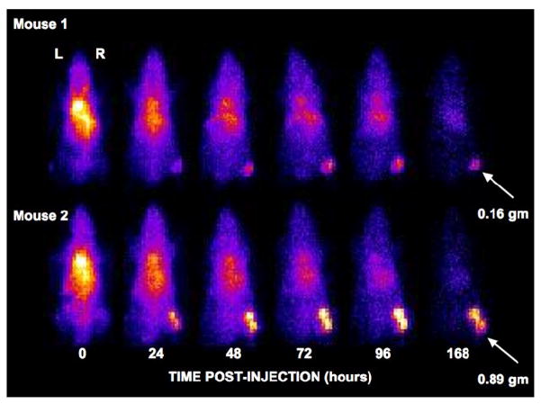
Methods: Two identical, miniature pixelated NaI(Tl) gamma cameras were fabricated and installed "looking up" through the tabletop of a compact portable cart. Mice are placed directly on the tabletop for imaging. Camera imaging performance was evaluated with phantoms and field performance was evaluated in a weeklong In-111 imaging study performed in a mouse tumor xenograft model.
Results: Tc-99m performance measurements, using a photopeak energy window of 140 keV+/-10%, yielded the following results: spatial resolution (FWHM at 1 cm), 2.2 mm; sensitivity, 149 cps (counts per seconds)/MBq (5.5 cps/microCi); energy resolution (FWHM, full width at half maximum), 10.8%; count rate linearity (count rate vs. activity), r(2)=0.99 for 0-185 MBq (0-5 mCi) in the field of view (FOV); spatial uniformity, <3% count rate variation across the FOV. Tumor and whole-body distributions of the In-111 agent were well visualized in all animals in 5-min images acquired throughout the 168-h study period.
Conclusion: Performance measurements indicate that MONICA is well suited to whole-body single photon mouse imaging. The field study suggests that inter-device communications and user-oriented interfaces included in the MONICA design facilitate use of the system in practice. We believe that MONICA may be particularly useful early in the (cancer) drug development cycle where basic whole-body biodistribution data can direct future development of the agent under study and where logistical factors, e.g., limited imaging space, portability and, potentially, cost are important.
Copyright 2010 Elsevier Inc. All rights reserved.
Figures
Similar articles
-
Performance characteristics of a positron projection imager for mouse whole-body imaging.Nucl Med Biol. 2013 Apr;40(3):321-30. doi: 10.1016/j.nucmedbio.2012.12.003. Epub 2013 Feb 9. Nucl Med Biol. 2013. PMID: 23402672 Free PMC article.
-
Performance characteristics of a new pixelated portable gamma camera.Med Phys. 2012 Jun;39(6):3435-44. doi: 10.1118/1.4718874. Med Phys. 2012. PMID: 22755723 Free PMC article.
-
Development of a pixelated GSO gamma camera system with tungsten parallel hole collimator for single photon imaging.Med Phys. 2012 Feb;39(2):581-8. doi: 10.1118/1.3673774. Med Phys. 2012. PMID: 22320767
-
Performance of a PSPMT based detector for scintimammography.Phys Med Biol. 2000 Mar;45(3):781-800. doi: 10.1088/0031-9155/45/3/315. Phys Med Biol. 2000. PMID: 10730971
-
Use of a compact pixellated gamma camera for small animal pinhole SPECT imaging.Ann Nucl Med. 2006 Jul;20(6):409-16. doi: 10.1007/BF03027376. Ann Nucl Med. 2006. PMID: 16922469
Cited by
-
In Vitro and In Vivo Pre-Clinical Analysis of a F(ab')(2) Fragment of Panitumumab for Molecular Imaging and Therapy of HER1 Positive Cancers.EJNMMI Res. 2011 Jun 7;1(1):1. doi: 10.1186/2191-219X-1-1. EJNMMI Res. 2011. PMID: 21845232 Free PMC article.
-
Performance characteristics of a positron projection imager for mouse whole-body imaging.Nucl Med Biol. 2013 Apr;40(3):321-30. doi: 10.1016/j.nucmedbio.2012.12.003. Epub 2013 Feb 9. Nucl Med Biol. 2013. PMID: 23402672 Free PMC article.
-
Characterization of "γ-Eye": a Low-Cost Benchtop Mouse-Sized Gamma Camera for Dynamic and Static Imaging Studies.Mol Imaging Biol. 2017 Jun;19(3):398-407. doi: 10.1007/s11307-016-1011-4. Mol Imaging Biol. 2017. PMID: 27730469
-
Performance evaluation of a newly developed high-resolution, dual-head animal SPECT system based on the NEMA NU1-2007 standard.J Appl Clin Med Phys. 2014 Nov 8;15(6):4936. doi: 10.1120/jacmp.v15i6.4936. J Appl Clin Med Phys. 2014. PMID: 25493518 Free PMC article.
-
SPECT detectors: the Anger Camera and beyond.Phys Med Biol. 2011 Sep 7;56(17):R145-82. doi: 10.1088/0031-9155/56/17/R01. Epub 2011 Aug 9. Phys Med Biol. 2011. PMID: 21828904 Free PMC article. Review.
References
-
- Proffitt J, Hammond W, Majewski S, et al. A Flexible High-rate USB2 Data Acquisition System for PET and SPECT Imaging. IEEE Nuclear Science Symposium Conference Record. 2005;5:2971–2975.
-
- Giusti RM, Shastri KA, Cohen MH, Keegan P, Pazdur R. FDA Drug Approval Summary: Panitumumab (Vectibix) Oncologist. 2007;12:577–583. - PubMed
-
- Loudos G, Majewski S, Wojcik R, et al. Performance Evaluation of a Dedicated Camera Suitable for Dynamic Radiopharmaceutical Evaluation in Small Animals. IEEE Transactions on Nuclear Science. 2007;50:454–460.
-
- MEDX, Inc; Arlington Heights, IL: T-Quest™ gamma camera product information. http://www.medx-inc.com/tquest.html.
Publication types
MeSH terms
Grants and funding
LinkOut - more resources
Full Text Sources
Medical
