

Comparative Study
doi: 10.1016/j.bbr.2010.03.029.
Epub 2010 Mar 25.
Deafening decreases neuronal incorporation in the zebra finch caudomedial nidopallium (NCM)
Affiliations
- PMID: 20346987
- PMCID: PMC2866162
- DOI: 10.1016/j.bbr.2010.03.029
Item in Clipboard
Comparative Study
Deafening decreases neuronal incorporation in the zebra finch caudomedial nidopallium (NCM)
Behav Brain Res.
.
Abstract
New neurons formed in the adult brain are incorporated into existing circuits. However, the number of new neurons recruited into a given brain region varies widely depending on the experience of the animal. An emerging general principle is that recruitment and early neuronal survival may be correlated with activity or use of the brain region. Here we show that use-dependent neuronal survival also occurs in the higher order auditory processing region of the songbird caudomedial nidopallium (NCM). We suggest that retention of young neurons may in part be influenced by use of the system without an increased demand for learning or behavioral plasticity.
Copyright 2010 Elsevier B.V. All rights reserved.
Figures
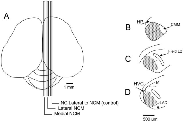
Labeled cells were compared in medial and lateral divisions of NCM, each containing 240 μm of tissue along the medial-lateral axis, separated by 336μm (A). A representative section in each division is shown as an outline drawn in Neurolucida using Lucivid: (B) Medial (~170–410 μm from midline), (C) Lateral (~746–986 μm from midline), (D) NC lateral to NCM (~1322–1562 from midline). Each tracing in NCM was divided into dorsal and ventral regions, approximately parallel to the dorsal and ventral edges of the brain and perpendicular to the caudal edge (dashed lines). Stippled areas indicate traced regions in which cells were counted. Anatomical landmarks: hippocampus (HP), caudal medial mesopallium (CMM), mesopallium (M), dorsal arcopallial lamina (LAD) arcopallium (A). Nucleus HVC and Field L2 are described in the text.
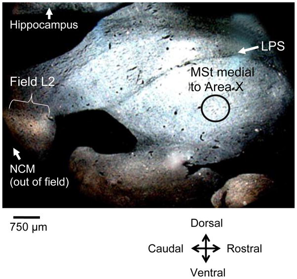
Darkfield rostroventral section showing the circular template (0.75 mm in diameter, black circle outline in figure) used to sample a control region of medial striatum (MSt) on 10 sections distributed approximately 200 μm-986 μm lateral from the midline. The template was placed ventral to the pallio-subpallialis lamina (LPS) and caudal to the area where Area X would emerge in more lateral sections. Field L is seen as a light band in the caudal region of the brain. NCM is caudal to Field L.
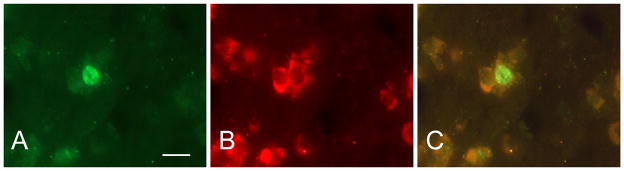
BrdU-labeled cells were identified with a fluorescein isothiocyanate (FITC) filter (A) and Hu-labeled cells were identified with a rhodamine filter (B). Double-labeled cells were identified by alternating between these two filters and also using a dual FITC-rhodamine filter (C). Scale bar = 20 μm
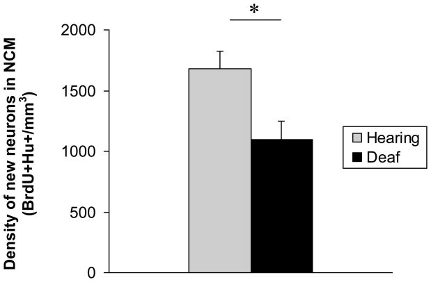
Deaf birds had a significantly lower density of new neurons in NCM than hearing birds.
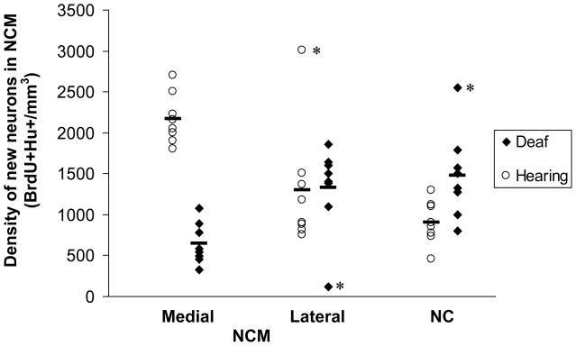
Deafening resulted in a significant decrease in new neuron density in medial NCM compared with hearing birds. There was no difference between deaf and hearing birds in new neuron density in lateral NCM or in caudomedial nidopallium (NC) lateral to NCM. Within the hearing group, the density of new neurons in the medial NCM was significantly higher than that in both the lateral NCM and NC. There was no difference in new neuron density between lateral NCM and NC in the hearing birds. Within the deaf group, new neuron density between the medial and lateral divisions was significantly different. Neither medial NCM nor lateral NCM were different from NC in the deaf birds. Removal of the 3 outliers (asterisks) did not change the results. Bars indicate means for each region within a treatment group, n=8 for each region within a treatment group.
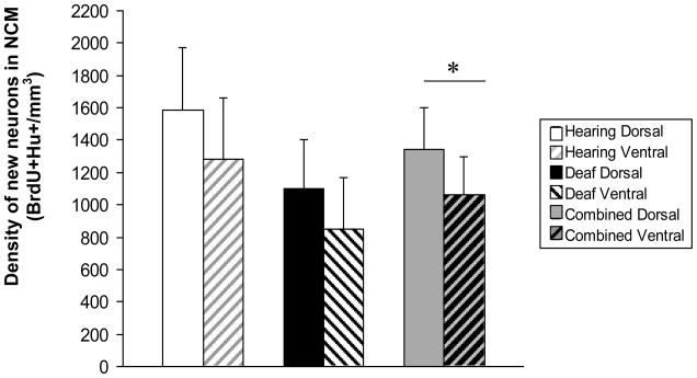
We found a significantly higher density of new neurons in dorsal NCM compared with ventral NCM when all birds were combined. The differences between dorsal and ventral NCM new neuron densities were not significant in either control or experimental groups considered separately, although the trend was similar in both groups.
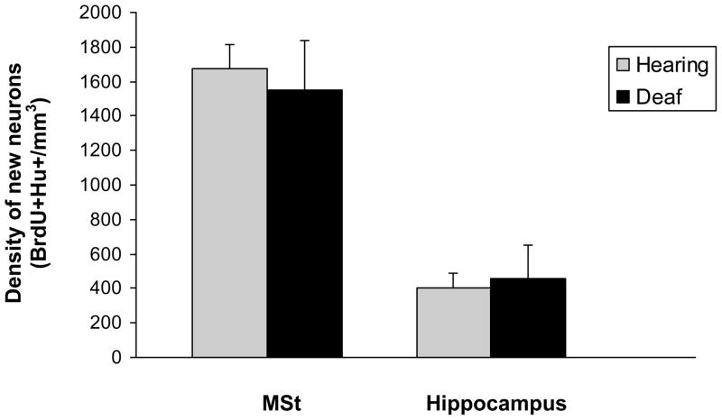
There was no difference in new neuron density in the medial striatum (MSt) or hippocampus between hearing and deaf birds.
References
-
- Adar A, Lotem A, Barnea A. The effect of social environment on singing behavior in the zebra finch (Taeniopygia guttata) and its implication for neuronal recruitment. Behav Brain Res. 2008;187:178–184. - PubMed
-
- Barnea A, Mishal A, Nottebohm F. Social and spatial changes induce multiple survival regimes for new neurons in two regions of the adult brain: An anatomical representation of time? Behav Brain Res. 2006;167:63–74. - PubMed
Publication types
MeSH terms
Grants and funding
LinkOut - more resources
Full Text Sources
