. 2010 Jul;31(19):5110-21.
doi: 10.1016/j.biomaterials.2010.03.003. Epub 2010 Mar 27.

The role of a recombinant fragment of laminin-332 in integrin alpha3beta1-dependent cell binding, spreading and migration

Affiliations

The role of a recombinant fragment of laminin-332 in integrin alpha3beta1-dependent cell binding, spreading and migration

Hironobu Yamashita et al. Biomaterials. 2010 Jul.

Abstract

The extracellular matrix (ECM) is thought to be an essential component of tissue scaffolding and engineering because it fulfills fundamental functions related to cell adhesion, migration, and three-dimensional organization. Natural ECM preparations, however, are challenging to work with because they are comprised of macromolecules that are large and insoluble in their functional state. Functional fragments of ECM macromolecules are a viable answer to this challenge, as demonstrated by the RGD-based engineered scaffolds, where the tri-peptide, Arg-Gly-Asp (RGD), represents the minimal functional unit of fibronectin and related ECM. Laminins (Ln) are main components of epithelial tissues, since they enter into the composition of basement membranes. Application of Ln to epithelial tissue engineering would be desirable, since they could help mimic ideal functional conditions for both lining and glandular epithelial tissues. However, functional fragments of Ln that could be used in artificial settings have not been characterized in detail. In this paper, we describe the production and application of the recombinant LG4 (rLG4) fragment of laminin-332 (Ln-332), and show that it mimics three fundamental functional properties of Ln-332: integrin-mediated cell adhesion, spreading, and migration. Adhesive structures formed by cells on rLG4 closely resemble those formed on Ln-332, as judged by microscopy-based analyses of their molecular composition. As on Ln-332, focal adhesion kinase (FAK) is phosphorylated in cells adhering to rLG4, and colocalized with other focal adhesion components. We conclude that rLG4 could be a useful substitute to recapitulate, in vitro, the tissue scaffolding properties of Ln-332.


Figures

Fig. 1
Fig. 1. Ln-332 structure and recombinant α3 LG4 expression in bacteria and mammalian cells
(A) Ln-332 is composed of α3, β3, and γ2 chains assembled into α3β3γ2 heterotrimer via a three-stranded α-helical coiled-coil domain. A globular domain (G domain) uniquely exists at the C-terminus of the α3 chain, which consists of 5 tandem laminin G-like (LG) repeats (LG1-LG5). (B) Schematic diagram of recombinant proteins. The recombinant α3 LG4 module was expressed using GST and His6 tag fusion protein in bacteria (BacLG4), and a His6 tagged protein in mammalian HEK-293 EBNA cells (MamLG4). GST alone and His6 tagged recombinant SAS alone were also expressed for control. (C) After GST and SAS constructs were purified and rLG4 modules were expressed in E. coli and Hek-293 cells, eluted material was subjected to SDS-PAGE analysis (NuPAGE 4 - 12% gel) under reducing conditions and visualized by Coomassie staining, which indicated proteins were purified to near homogeneity since they show up as single bands. (D) Immunoblot analysis was carried out using an mAb against His6 tag, which detected rLG4 proteins and SAS, but not GST due to its lack of this structure (D).
Fig. 2
Fig. 2. Mass spectrometry analysis of bacterial and mammalian rLG4
After in-gel trypsin digestion of rLG4 modules, peptides were identified by MALDI-TOF mass spectrometry. (A) Mass spectrometry identified 9 different peptides in BacLG4 digesion. (B) Localization of peptides (* 1-9) for BacLG4 detected by mass spectrometry was compared with the established rat Ln-332 α3 chain sequence (1312-1552+His6 tag). Labels A-M signify the folds in the β sheet structure. (C) Mass spectrometry identified 9 different peptides in MamLG4 digesion. (D) Localization of peptides (*1-9) for MamLG4 was compared with the established rat laminin-332 α3 chain sequence (1312-1552+His6 tag). Labels A-M signify the folds in the β sheet structure.
Fig. 3
Fig. 3. HT-1080, A-431, and keratinocytes adhere to rLG4
96-well plates were coated at 4 °C overnight with BacLG4, GST protein, MamLG4, SAS, or Ln-332. A-431, HT-1080, or keratinocytes were seeded and incubated on substrates for 1-2 h at 37 °C. Non-adherent cells were then washed from wells and remaining cells were stained with CV, and absorbance was measured as indicated. Results are expressed as the mean absorbance ± standard deviation. (A, B) Both A-431 and HT-1080 lines adhered to BacLG4 and MamLG4, similarly to intact Ln-332 (N = 3; P = 0.016-0.91). In contrast, GST and SAS controls had almost no adhesion activity (N = 3; P < 0.001). (C) Similarly, human keratinocytes adhered to MamLG4 similarly to Ln-332 (N = 2; P < 0.001).
Fig. 4
Fig. 4. Cell adhesion to rLG4 is integrin-dependent
(A) In adhesion experiments involving the use of EDTA, 96-well plates were coated at 4 °C overnight with BacLG4, MamLG4, or Ln-332 protein. Where indicated, A-431 and HT-1080 cells were treated with EDTA for 1 h prior to plating. When EDTA was added to either cell line prior to incubation, adhesion was significantly reduced on all substrates (N=3; P < 0.001). (B) In Mn2+ experiments, 96-well plates were coated at 4 °C overnight with BacLG4, MamLG4, Ln-332, GST, or SAS protein. Where indicated, cells were treated with Mn2+ at various concentrations for 1 h at 37 °C. Cells were seeded (, and allowed to adhere to substrates for 1 h at 37°C. Non-adherent cells were washed off, adherent cells were stained with CV, and absorbance was measured at 570 nm. Results are presented as the mean absorbance ± standard deviation (N=3). Stimulation of cells with Mn2+ significantly enhanced the number of cells that adhered to BacLG4, MamLG4, and Ln-322 (P<0.05, in all cases), but not GST or SAS. Taken together, these results indicate that rLG4 stimulates binding activity in an integrin-dependent manner.
Fig. 5
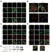
Fig. 5. rLG4 induces cell spreading and focal adhesion structures
(A) A431 cells were seeded on glass coverslips coated with BacLG4, MamLG4, Ln-332, SAS, or GST, and allowed to adhere for 1 h at 37°C. After a washing step, adherent cells were labeled with Hoechst to stain nuclei (blue), phalloidin to locate actin organization (green), and paxillin to target focal contacts (red). Cells plated on MamLG4 and BacLG4 exhibited cell spreading, actin organization, and focal adhesion formation similar to cells on Ln-332 substrate. (B) Human epidermal keratinocytes were also seeded on glass dishes coated with Ln-332, MamLG4, SAS, or PBS, and allowed to adhere for 2 h at 37°C. After washing, adherent cells were labeled with Hoechst (blue) and anti-paxillin (primary mAb), followed by secondary IgG Alexa Fluor 568 (red) and Alexa Fluor 488 (green) to detect focal adhesions and F-actin, respectively. Keratinocytes plated on MamLG4 exhibited cell spreading, actin organization, and focal adhesion formation similar to cells on Ln-332 substrate. Whereas cells plated on SAS or PBS exhibited little organization or spreading. (C) A-431 cells were handled similarly to part A, except cells were stained for p-FAK to identify tyrosine phosphorylation (red), in lieu of paxillin. This marker revealed that FAK is phosphorylated similarly in on both Ln-332 and rLG4. (D) Similarly, keratinocytes spread and showed phosphorylated FAK on MamLG4 and Ln-332, but not on SAS or PBS. Highly magnified regions of interest highlight the colocalization of paxillin and pFAK markers, which indicates that phosphorylation occurs at sites of focal adhesion. (E) Western blotting analysis confirms that tyrosine phosphorylation of FAK was induced in A-431 cells attached to immobilized rLG4 (similarly to Ln-332), in contrast to cells on SAS or PBS.
Fig. 6
Fig. 6. Integrin α3β1 mediates cell adhesion and migration on rLG4
Adhesion (A) and haptotactic migration (B) activity of HT-1080 and A-431 cell lines against rLG4 were measured using standard 96-well plate adhesion and migration assays. Where indicated, cells were pre-incubated with EDTA or the following inhibitory antibodies: P1B5 (anti-α3), 6S6 (anti-β1), P1D6 (anti-α5), or GoH3 (anti-α6) for 30 min. Plots represent the mean ± standard deviation percentage of adhesion (N=3) or migration (N=2) relative to untreated cells set to 100% (on BacLG4 or MamLG4). Both cell adhesion and/or migration were significantly inhibited by anti-α3, anti-β1, and EDTA (P < 0.05, in all cases), but not by anti-α6 or anti-α5 antibodies. These results indicate that cell adhesion and migration on rLG4 is dependent on α3β 1 integrin.
Fig. 7
Fig. 7. Integrin β1-knockdown decreases cell spreading on rLG4
(A) IMCD-WT and IMCD-β1 null cells were seeded and allowed to adhere for 1 h at 37 °C. After a wash step, bound cells were stained with CV, and microphotographs were taken at 400× magnification. Values represent the mean ± standard deviation percentage of spread cells relative to total cell number set to 100% (N=3). (B) β1-IMCD null and WT cells were plated on Ln-332 or MamLG4 and allowed to adhere for 1 h. After a wash step, adherent cells were labeled with Hoechst stain nuclei (blue), phalloidin to locate actin organization (red), and α3 integrin pAb 1948 (green). Microphotographs were taken at 630× magnification.
Fig. 8
Fig. 8. Integrin α3β1 specifically binds to rLG4 module
GST pull-down assays were performed to obtain direct evidence for interaction of rLG4 with α3β1 integrin. Assays used BacLG4 and HT-1080 cell lysates. GSH-sepharose bound GST or BacLG4 was incubated with HT-1080 cell lysates in the presence of Mn2+ ion. After incubation, beads were centrifuged at 5,000 rpm for 5 min and washed 3 times with 5 bed volume of lysis buffer. (A) Bound material was mixed with non-reducing sample buffer for SDS-PAGE and boiled for 5 min at 100 °C. (B) Western blot was carried out with antibodies against integrin α3 or β 1. These results indicate α3β 1 binds specifically to rLG4.

Similar articles

Cited by

References

    1. Rozario T, Desimone DW. The extracellular matrix in development and morphogenesis: A dynamic view. Dev Biol. 2009 Oct 23; Epub ahead of print. - PMC - PubMed
    1. Menko AS, Boettinger D. Occupation of the extracellular matrix receptor integrin is a control point for differentiation. Cell. 1987;51:51–7. - PubMed
    1. Zhu X, Ohtsubo M, Bohmer RM, Roberts JM, Assoian RK. Adhesion-dependent cell cycle progression linked to the expression of cyclin D1, activation of cyclin E-cdk2, and phosphorylation of the retinoblastoma protein. J Cell Biol. 1996;133:391–403. - PMC - PubMed
    1. Rosso F, Giordano A, Barbarisi M, Barbarisi A. From cell-ECM interactions to tissue engineering. J Cell Physiol. 2004;119(2):174–80. - PubMed
    1. Hynes RO. Integrins: versatility, modulation, and signaling in cell adhesion. Cell. 1992;69:11–25. - PubMed

Publication types

MeSH terms