. 2010 May;9(3):217-27.
doi: 10.1016/j.jcf.2010.03.001. Epub 2010 Mar 29.

Alveolar inflammation in cystic fibrosis

Affiliations

Alveolar inflammation in cystic fibrosis

Martina Ulrich et al. J Cyst Fibros. 2010 May.

Abstract

Background: In infected lungs of the cystic fibrosis (CF) patients, opportunistic pathogens and mutated cystic fibrosis transmembrane conductance regulator protein (CFTR) contribute to chronic airway inflammation that is characterized by neutrophil/macrophage infiltration, cytokine release and ceramide accumulation. We sought to investigate CF lung inflammation in the alveoli.

Methods: Lung tissue from 14 CF patients and four healthy individuals was analyzed for numbers of effector cells, elastin and collagen concentrations, inflammatory markers and density of Pseudomonas aeruginosa. Additionally, desmosine and isodesmosine concentrations were determined in 52 urine specimens from CF patients to estimate the burden of elastase activities in respiratory secretions.

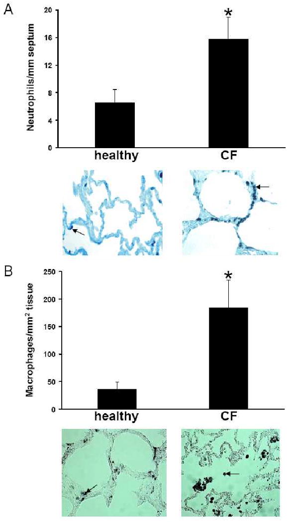
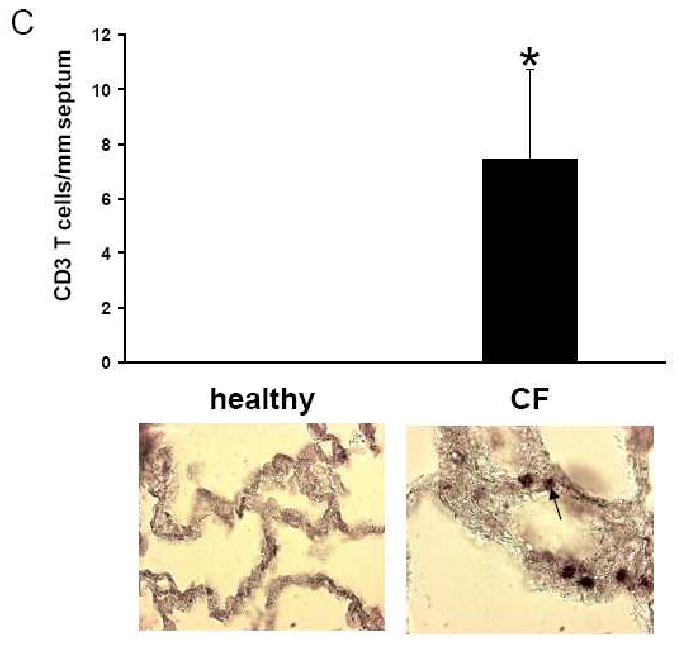
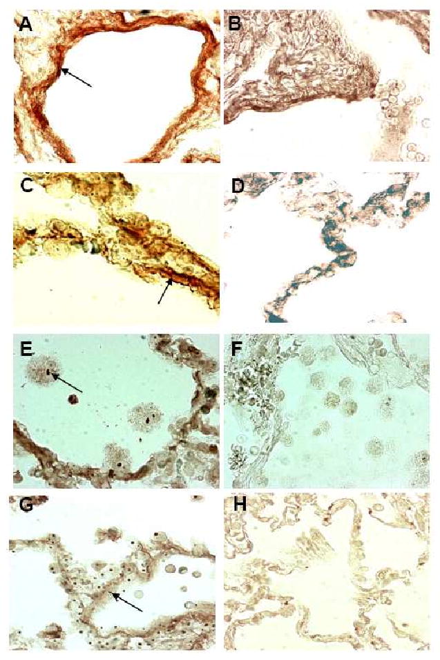
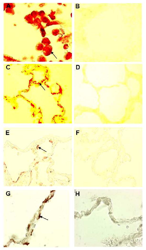
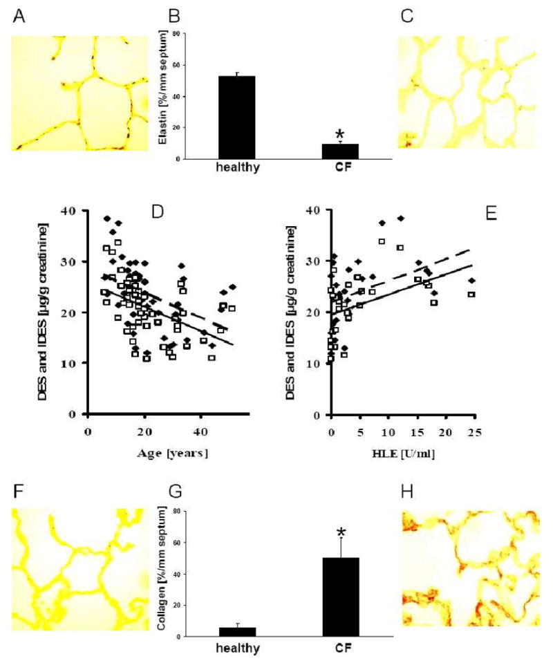
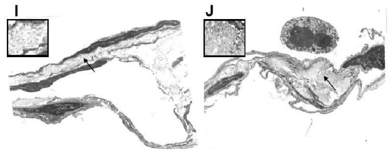
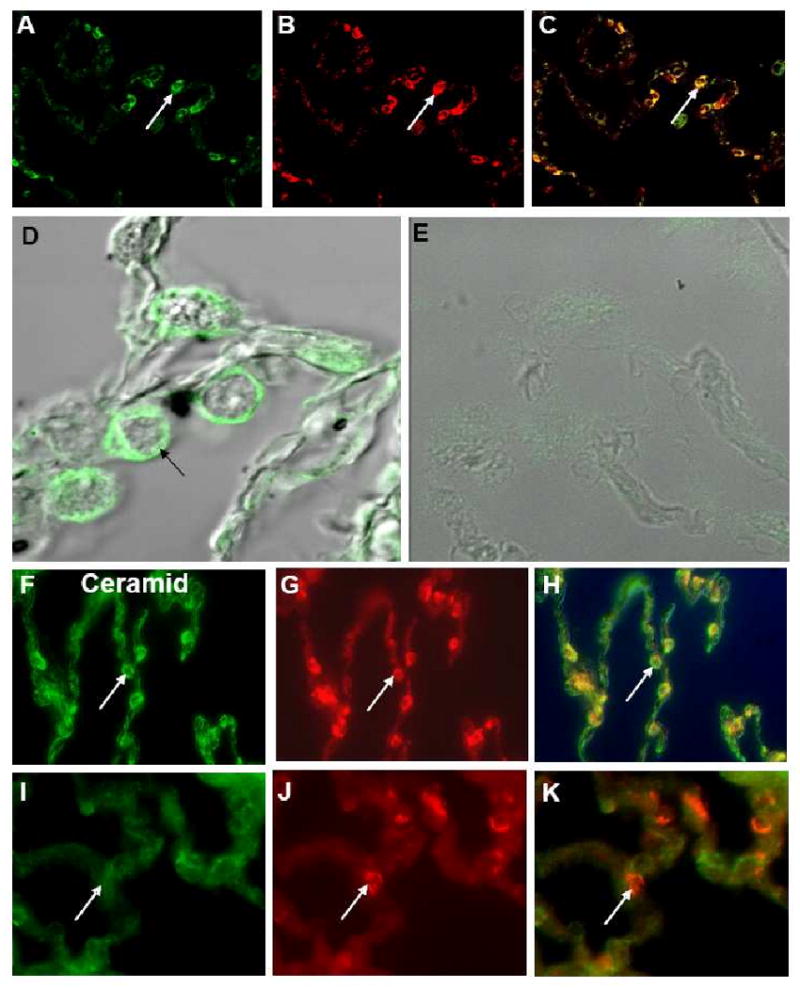
Results: Elastin concentration was significantly decreased and collagen significantly increased in CF alveolar tissues as compared to age-matched, healthy individuals. Elastin split products were significantly increased in urine samples from patients with CF and correlated inversely with age, indicating local tissue remodelling due to elastin degradation by unopposed proteolytic enzymes. Alveolar inflammation was also characterized by a significant cell infiltration of neutrophils, macrophages and T cells, extensive nuclear factor-kappaB and insulin-like growth factor-1 activation in various cell types and increased intercellular adhesion molecule-1 expression, and increased numbers of myofibroblasts. Additionally, ceramide accumulated in type II alveolar epithelial cells, lacking CFTR. P. aeruginosa organisms were rarely present in inflamed alveoli.

Conclusions: Chronic inflammation and remodeling is present in alveolar tissues of the CF lung and needs to be addressed by anti-inflammatory therapies.


Figures

Figure 1
Figure 1. Increased numbers of neutrophils, alveolar macrophages and T lymphocytes are present in alveoli of CF patients
Neutrophils (A, arrow), stained with monoclonal antibodies directed against human neutrophil elastase, in CF alveolar lung tissue and in normal alveolar lung tissue differ significantly (p<0.0001) in numbers. Alveolar macrophages (B, arrow), stained with monoclonal antibodies directed against CD68 receptors, in CF alveolar lung tissue and in normal alveolar lung tissue differ significantly (p<0.0001) in numbers with regard to cells in septa (black bars) and intraluminally (white bars). T lymphocytes (C, arrow), stained with monoclonal antibodies directed against CD3, in CF alveolar lung tissue and in normal alveolar lung tissue differ significantly (p<0.0001) in numbers.
Figure 2
Figure 2. Inflammatory mediators and transcription factor expression in CF versus normal alveolar lung tissue
Immunohistochemical detection of ICAM-1 expression in alveolar tissue of a CF patient (A, C) and a healthy individual (B, D). In the CF patient, positively stained (dark brown) ICAM-1 expression was present in blood vessels (A, arrow) and alveolar septae (C, arrow), whereas blood vessels (B) and alveolar septae (D) of a healthy individual were negative. E, G: Immunohistochemical detection of NF-κB in the alveolar tissue of a CF patient. All alveolar macrophages in alveolar lumina (E) and many cells in the alveolar epithelium (G) of CF lungs were positive for NF-κB staining in the nuclei (dark brown spots, arrow). These cells were negative for nuclear NFκB in a healthy control (F, H). Original magnifications: D-J: × 200
Figure 3
Figure 3. IGF-1 and myofibroblast expression in CF versus normal alveolar lung tissue
IGF-1, stained with a polyclonal antibody (red), differed significantly (p<0.0001) between CF alveolar (A, C) and normal alveolar lung tissue (B, D). Myofibroblasts, stained with an antibody (dark brown) in CF alveolar lung tissue (E, G) and in normal alveolar lung tissue (F, H) differed largely in expression. Original magnifications: A, B, F, H × 400; C, D, E, G × 200.
Figure 4
Figure 4. Increased elastin degradation and collagen substitution in alveolar septa and increased elastin degradation in urines of CF patients
Elastin fibers, stained with a rabbit IgG antibody directed against human α-elastin, in CF alveolar lung tissue (C) and in normal alveolar lung tissue (A) differ significantly (p<0.0001) in concentration (B). In CF patients mean elastin values ± SD were 9.6±1.5% elastin/mm septum (range: 7.6-12.6% elastin/mm septum), and 52.5±2.3% elastin/mm septum (range: 50.3-55.1% elastin/mm septum) in healthy control lungs. Urinary concentrations of the elastin split product desmosine (DES □) and isodesmosine (IDES ◆), determined by micellar electrokinetic chromatography, decrease with age in 52 CF patients (D). Both markers of elastin degradation were highly correlated. DES and IDES concentrations correlate with human neutrophil elastase (HLE) levels in sputum samples of CF patients (E). Collagen fibers, stained with a rabbit IgG antibody directed against human collagen type 1, in CF alveolar lung tissue (H) and in normal alveolar lung tissue (F) differ significantly (p<0.0001) in concentration (G). Mean collagen type 1 expression ± SD in alveolar septa of CF patients was 49.9±13.2% collagen/mm septum (range: 30.2-74.6% collagen/mm septum) and 7.3±2.6% collagen/mm septum, range: 4.0-9.6% collagen/mm septum) in tissues of healthy controls. Normal collagen deposition in alveolar septae of a healthy individual (I) and increased deposition of collagen in alveolar septae of a CF patient (J). Arrows: Insert. Original magnifications: A, C, G, I × 100; Original magnifications: A, C, F, H × 100; I, J × 3.190.
Figure 5
Figure 5. Ceramide accumulates in type II alveolar epithelial cells of CF patients
Immunostaining of alveolar type II cells from healthy individuals with a monoclonal antibody specific for CFTR (A) and a rabbit antibody against surfactant C (B); C: overlay of A and B. D, E: Confocal microscopy of alveolar type II cells from a healthy individual (D) and a ΔF508 homozygous CF patient. (E) with a monoclonal antibody specific for CFTR. Immunostaining of alveolar type II cells from CF patients (F, G) and healthy individuals (I, J) with a monoclonal antibody specific for ceramide (F, I) and a rabbit antibody against surfactant C (G, J). H: overlay of F, G, K: overlay of I,J. Original magnifications: A-C, F-K: × 200; D, E × 400.

Similar articles

Cited by

References

    1. Rommens JM, Iannuzzi MC, Kerem B, Drumm ML, Melmer G, Dean M, Rozmahel R, Cole JL, Kennedy D, Hidaka N, Zsiga M, Buchwald M, Riordan JR, Tsui LC, Collins F. Identification of the cystic fibrosis gene: chromosome walking and jumping. Science. 1989;245:1059–65. - PubMed
    1. Riordan JR, Rommens JM, Kerem B, Alon N, Rozmahel R, Grzelczak Z, Zielenski J, Lok S, Plavsic N, Chou JL, Drumm ML, Iannuzzu MC, Collins FS, Tsui LC. Identification of the cystic fibrosis gene: cloning and characterization of complementary DNA. Science. 1989;245:1066–73. - PubMed
    1. Kerem B, Rommens JM, Buchanan JA, Markiewicz D, Cox TK, Chakravarti A, Buchwald M, Tsui LC. Identification of the cystic fibrosis gene: genetic analysis. Science. 1989;245:1073–80. - PubMed
    1. Boucher RC. New concepts of the pathogenesis of cystic fibrosis lung disease. Eur Respir J. 2004;23:146–58. - PubMed
    1. Worlitzsch D, Tarran R, Ulrich M, Schwab U, Cekici A, Meyer KC, Birrer P, Bellon G, Berger J, Weiß T, Botzenhart K, Yankaskas JR, Randell S, Boucher RC, Döring G. Reduced oxygen concentrations in airway mucus contribute to the early and late pathogenesis of Pseudomonas aeruginosa cystic fibrosis airways infection. J Clin Invest. 2002;109:295–09. - PMC - PubMed

MeSH terms