. 2010 Mar 26;37(6):797-808.
doi: 10.1016/j.molcel.2010.02.022.

Postnatal deamidation of 4E-BP2 in brain enhances its association with raptor and alters kinetics of excitatory synaptic transmission

Affiliations

Postnatal deamidation of 4E-BP2 in brain enhances its association with raptor and alters kinetics of excitatory synaptic transmission

Michael Bidinosti et al. Mol Cell. .

Abstract

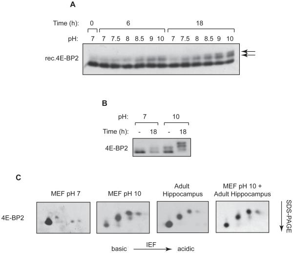
The eIF4E-binding proteins (4E-BPs) repress translation initiation by preventing eIF4F complex formation. Of the three mammalian 4E-BPs, only 4E-BP2 is enriched in the mammalian brain and plays an important role in synaptic plasticity and learning and memory formation. Here we describe asparagine deamidation as a brain-specific posttranslational modification of 4E-BP2. Deamidation is the spontaneous conversion of asparagines to aspartates. Two deamidation sites were mapped to an asparagine-rich sequence unique to 4E-BP2. Deamidated 4E-BP2 exhibits increased binding to the mammalian target of rapamycin (mTOR)-binding protein raptor, which effects its reduced association with eIF4E. 4E-BP2 deamidation occurs during postnatal development, concomitant with the attenuation of the activity of the PI3K-Akt-mTOR signaling pathway. Expression of deamidated 4E-BP2 in 4E-BP2(-/-) neurons yielded mEPSCs exhibiting increased charge transfer with slower rise and decay kinetics relative to the wild-type form. 4E-BP2 deamidation may represent a compensatory mechanism for the developmental reduction of PI3K-Akt-mTOR signaling.


Figures

Figure 1
Figure 1. Slow-migrating forms of 4E-BP2 in the adult brain are independent of phosphorylation
(A) Reduced phosphorylation and a slow-migrating 4E-BP2 form are unique to the brain. Mouse tissue lysates (3 months) were analyzed by Western blotting (15% SDS-PAGE). (B) Far Western binding of eIF4E to 4E-BP2 from brain. Brain lysates were resolved and probed on a nitrocellulose membrane with radiolabeled FLAG-HMK-eIF4E protein. Recombinant 4E-BP1 and a variant lacking the eIF4E-binding site (Δ51-63) control for probe specificity. Western blotting was with antibody #11211. (C) 4E-BP2 from mouse brain is insensitive to phosphatase treatment. Brain lysates were incubated with λ phosphatase followed by Western blotting. phospho-Thr-Pro motif antibody demonstrates phosphatase efficacy (lower panel). See also Figure S1.
Figure 2
Figure 2. 4E-BP2 is susceptible to asparagine deamidation in the mammalian brain
(A) Deamidation of recombinant 4E-BP2 at high pH. Recombinant 4E-BP2 was incubated in 0.15M Tris-HCl at the indicated pH and times, followed by Western blotting. Arrow heads indicate deamidated forms of 4E-BP2. (B) Deamidation of 4E-BP2 in cell lysates. MEFs were starved of serum for 24 hours, followed by one hour in PBS. Cells were lysed at the indicated pH and extracts were incubated for 18 hours. (C) 4E-BP2 deamidation caused by alkaline treatment or occurring spontaneously in hippocampus exhibits identical migration pattern in two-dimensional isoelectric focusing/SDS-PAGE (IEF/SDS-PAGE). Alkaline-treated MEF or untreated hippocampal lysates were analysed (panels 1-3). In panel 4, a 1:1 mixture of pH 10-treated MEF and hippocampal lysates was analysed. See also Figure S2.
Figure 3
Figure 3. Deamidation of 4E-BP2 occurs on asparagines 99 and 102
(A) Amino acid sequence alignment of murine 4E-BP2 and 4E-BP1. Inset shows the asparagine-rich sequence of 4E-BP2. (B) 4E-BP2 is sensitive to alkaline-induced deamidation. MEFs were lysed and incubated as in (Figure 2B). (C) A 4E-BP2 variant lacking the asparagine-rich sequence is resistant to deamidation. HEK293E cells were transfected with N-terminal 3HA-tagged 4E-BP2 or a variant lacking residues 87-104. Post-transfection (24 hours) cells were lysed at pH 7 or 10 and incubated as indicated. (D) Asn99 and Asn102 are favoured for deamidation. Purified, recombinant 4E-BP2 protein was in vitro deamidated at pH 10 for 24h, resolved by SDS-PAGE (10-20%), and stained (left panel). Excised bands were analysed by LC-MS/MS (right). (E) Mutation of both Asn99 and Asn102 to alanines precludes alkaline-induced deamidation. HEK293E cells were transfected and treated as in (C). (F) Mutation of Asn99 and Asn102 to aspartates retards 4E-BP2 migration. Cells were transfected as in (E). See also Figure S3.
Figure 4
Figure 4. Deamidated 4E-BP2 exhibits increased raptor-interaction, reduced association to eIF4E and attenuated translational repression
(A) Deamidated 4E-BP2 exhibits enhanced affinity for raptor. HEK293 cells were co-transfected with Myc-tagged raptor and 3HA-tagged 4E-BP2. Anti-Myc immunoprecipitation was performed on lysates 24 hours later, followed by Western blotting (7-15% SDS-PAGE). (B) Deamidated 4E-BP2 is weakly associated to eIF4E in cells grown in complete medium. Lysates were incubated with m7GDP agarose and bound proteins were analyzed by Western blotting. (C and D) Deamidated 4E-BP2 is more weakly associated to eIF4E in the absence of serum or nutrients. Cells were starved of serum for 24 hours (C) or incubated in D-PBS for 1 hour (D) followed by m7GDP pulldown. (E) m7GDP pulldowns from brain lysates were performed as in (B). (F) Deamidation of 4E-BP2 does not affect eIF4E-binding in the absence of raptor. Far Western blotting was performed as in Figure 1C against recombinant proteins. (G and H) Deamidated 4E-BP2 weakly inhibits translation. 4E-BP1/4E-BP2 null MEFs were co-transfected with 4E-BP2 together with IRF7-Firefly Luciferase and Renilla luciferase plasmids (G) or cultured neurons were co-transfected with 4E-BP2 plasmids and a reporter encoding myrdYFP flanked by the 5′ and 3′ UTRs of CamKIIα. MEFs in (G) were harvested 24 hours post-transfection and luciferase activity was measured (data are means ± SEM, with statistical significance set at p < 0.05). Cultured neurons in (H) were transfected 10 days after plating and two days later images of YFP-expressing neurons were acquired (N=9-12 cells; bars represent means ± SEM and in all cases, except amongst anisomycin treatments, means are different at p < 0.05 by Student’s t-test). 4E-BP2 expression was determined by immunofluorescence and no differences between wild type and deamidated 4E-BP2 expression was observed in proximal dendrites (not shown). See also Figure S4.
Figure 5
Figure 5. Deamidation of 4E-BP2 occurs during early postnatal brain development, concomitant with reduced PI3K-Akt-mTOR signaling
(A) Western blot analysis on hippocampal extracts (30μg each) from several postnatal (P) days compared with adult. (B) IEF/SDS-PAGE analysis of P3 hippocampal extract. (C) IEF/SDS-PAGE analysis of hippocampal extracts from indicated postnatal days. (D) Phosphatase treatment of P15 hippocampal extract. (E) SDS-PAGE analysis of lysates from dissociated hippocampal neuron cultures or brain from indicated days. (F) Analysis of associated eIF4E-binding proteins during hippocampal neuron development by m7GDP precipitation. See also Figure S5.
Figure 6
Figure 6. Expression of deamidated 4E-BP2 in 4E-BP2−/− neurons reduces elevated excitatory synaptic transmission but slows kinetics of mEPSCs
(A) Increased excitatory miniature synaptic activity in slices from 4E-BP2−/− mice. Left: Traces of mEPSCs from 4EBP2−/− and wild type mice. Right: Cumulative probability plots and summary bar graph for all cells showing increases in mEPSC amplitude, charge and frequency. (N=6 neurons/genotype and 200 events/neuron; data are means ± SEM. p < 0.05. Kolmogorov-Smirnov test). (B) Expression of deamidated or wild type 4E-BP2 in 4E-BP2−/− neurons. Left: mEPSC traces from 4E-BP2−/− organotypic slices transfected with empty vector (vec), wildtype 4E-BP2, or deamidated 4E-BP2 (N99D/N102D). Transfected cells were identified by IRES-mediated eGFP expression from the same plasmid encoding 4E-BP2 variants. Right top: Cumulative probability plots of mEPSC amplitude, charge, and frequency. Right bottom: Summary of effects on mEPSC amplitude, charge and frequency. (N=6 neurons/condition and 150 events/neuron; * p < 0.05, ** p < 0.01. One-way ANOVA). (C) Partial repression of mEPSC charge by deamidated 4E-BP2 is associated with slower kinetics. Top: Individual (10 traces, grey) and averaged (150 events, black) mEPSCs from neurons transfected as in (B). Superimposed scaled responses of average mEPSCs, are at the right. Bottom left: Cumulative probability plots for 10-90% rise and 90-10% decay times for all groups (p < 0.05, Kolmogorov-Smirnov test). Bottom right: Summary bar graphs of rise and decay times. (N=6, p < 0.01. One-way ANOVA). See also Figure S6.

Similar articles

Cited by

References

    1. Aakalu G, Smith WB, Nguyen N, Jiang C, Schuman EM. Dynamic visualization of local protein synthesis in hippocampal neurons. Neuron. 2001;30:489–502. - PubMed
    1. Banko JL, Hou L, Poulin F, Sonenberg N, Klann E. Regulation of eukaryotic initiation factor 4E by converging signaling pathways during metabotropic glutamate receptor-dependent long-term depression. J Neurosci. 2006;26:2167–2173. - PMC - PubMed
    1. Banko JL, Merhav M, Stern E, Sonenberg N, Rosenblum K, Klann E. Behavioral alterations in mice lacking the translation repressor 4E-BP2. Neurobiol Learn Mem. 2007;87:248–256. - PubMed
    1. Banko JL, Poulin F, Hou L, DeMaria CT, Sonenberg N, Klann E. The translation repressor 4E-BP2 is critical for eIF4F complex formation, synaptic plasticity, and memory in the hippocampus. J Neurosci. 2005;25:9581–9590. - PMC - PubMed
    1. Bekkers JM, Stevens CF. Cable properties of cultured hippocampal neurons determined from sucrose-evoked miniature EPSCs. J Neurophysiol. 1996;75:1250–1255. - PubMed

Publication types

MeSH terms

Substances