. 2010 Mar 26;37(6):834-42.
doi: 10.1016/j.molcel.2010.01.037.

The histone chaperone Nap1 promotes nucleosome assembly by eliminating nonnucleosomal histone DNA interactions

Affiliations

The histone chaperone Nap1 promotes nucleosome assembly by eliminating nonnucleosomal histone DNA interactions

Andrew J Andrews et al. Mol Cell. .

Abstract

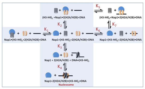
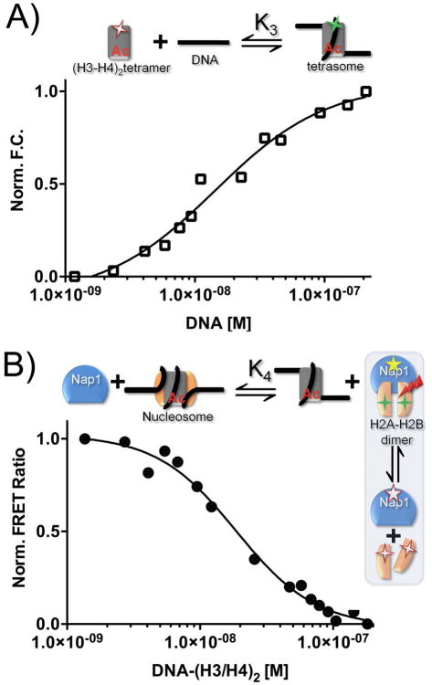
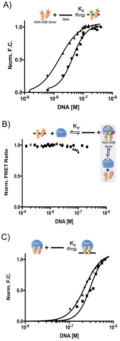
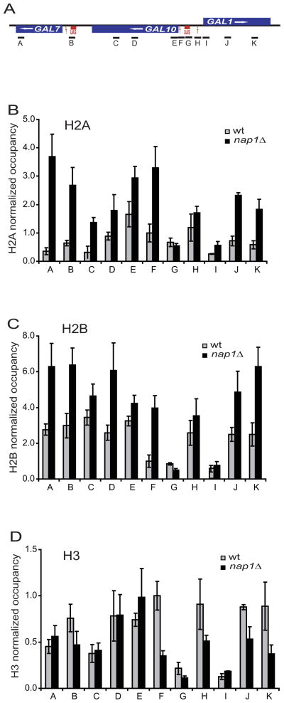
The organization of the eukaryotic genome into nucleosomes dramatically affects the regulation of gene expression. The delicate balance between transcription and DNA compaction relies heavily on nucleosome dynamics. Surprisingly, little is known about the free energy required to assemble these large macromolecular complexes and maintain them under physiological conditions. Here, we describe the thermodynamic parameters that drive nucleosome formation in vitro. To demonstrate the versatility of our approach, we test the effect of DNA sequence and H3K56 acetylation on nucleosome thermodynamics. Furthermore, our studies reveal the mechanism of action of the histone chaperone nucleosome assembly protein 1 (Nap1). We present evidence for a paradigm in which nucleosome assembly requires the elimination of competing, nonnucleosomal histone-DNA interactions by Nap1. This observation is confirmed in vivo, wherein deletion of the NAP1 gene in yeast results in a significant increase in atypical histone-DNA complexes, as well as in deregulated transcription activation and repression.


Figures

Figure 1
Figure 1. Thermodynamic scheme for Nap1 mediated nucleosome formation
Nap1 is shown in blue, (H3–H4)2 tetramer in grey, and H2A–H2B dimer in orange. DNA is shown as a black line. The shaded area depicts a mechanism in which simple competition between Nap1 and DNA for histones is sufficient for nucleosome assembly. This scheme is extended by adding the ternary Nap1-H2A–H2B-DNA complex. Equilibrium constants K1–K6 were measured as described in Figures 2–4, and are listed in Table 1. K7 was calculated. See supplementary material for details.
Figure 2
Figure 2. Measurement of the thermodynamic constants for nucleosome formation
The experimental design for each reaction is shown above each panel, using the symbols described in Fig. 1. Fluorescent labels are indicated by asterisks; FRET is indicated by a red arrow. Closed squares are 601 DNA or (H3–H4)2 tetramer-bound 601 sequence (601-tetrasomes); closed triangles are 5S sequence DNA or (H3–H4)2 tetramer-bound 5S DNA (5S tetrasomes). (A) Normalized fluorescence as a function of DNA binding to H3–H4. (B) The change in FRET between Nap1 and H2B as a function of tetrasome ((H3–H4)2-DNA). (C) The change in FRET between Nap1 and H2B as a function of (H3–H4)2. The Nap1 concentration is kept at the Kd for the Nap1-H2A–H2B dimer complex (K2, Table 1).
Figure 3
Figure 3. H3K56ac destabilizes tetrasome formation
Fluorescence studies of H3K56ac-H4 in tetrasome and nucleosome formation. (A) tetrasome formation as monitored by the normalized fluorescence change from Alexa-488 labeled K3K56ac-H4 as a function of DNA. (B) nucleosome assembly, as monitored by the FRET ratio between Nap1 and H2A–H2B as a function of H3K56ac-H4. Thermodynamic constants are shown in Table 1, experimental details are found in the supplemental section.
Figure 4
Figure 4. Nap1 disfavors the interaction between H2A–H2B dimer and DNA
The experimental design for each reaction is shown above each panel, using the symbols described in Fig. 1. Fluorescent labels are indicated by asterisks; FRET is indicated by a red arrow. Closed squares are 601-tetrasomes; closed triangles are 5S-tetrasomes. (A) Normalized fluorescence as a function of DNA binding to H2A–H2B. (B) The change in FRET between Nap1 and H2B as a function of DNA. The experiment was originally designed to confirm K6; however, the data indicate the Nap1-H2A–H2B complex remains intact in the presence of DNA and thus the small amount of signal change is due to DNA binding to the Nap1-H2A–H2B complex. Therefore, the binding event really monitored is K5, and is thus termed K5*. (C) Fluorescence change as a function of DNA binding to a Nap1-H2A–H2B complex (Nap1 is 10-fold > K2). Binding constants derived from the shown experiments are summarized in Table 1. See supplemental information for experimental details.
Figure 5
Figure 5. Deletion of NAP1 alters histone occupancy in vivo
(A) schematic of the GAL gene locus, showing the relative position of the coding sequence for GAL1, GAL7 and GAL10. The location of the amplicons used for ChIP assays are indicated (A–K). (B) ChIP analyses of histone H2A were performed on wild type (wt; grey bars) or nap1Δ cells (black bars). Each column corresponds to the location of a real-time PCR amplicon as shown in (A). Error bars indicate standard deviations from three independent biological replicates. Histone H2B (C) and H3 (D) occupancies were determined as for H2A.
Figure 6
Figure 6. Deletion of NAP1 changes the kinetics of GAL gene expression
(A) S1 nuclease protection assays were used to analyze the expression levels of GAL1 and GAL7 transcripts in a wild type or nap1Δ strain. To examine activation of transcription, galactose was added for the time (in minutes) indicated. To examine repression of transcription, glucose was added after 60 minutes of growth in galactose. A representative gel is shown. (B, C) The amount of transcript for GAL1 and GAL7 at each time point is plotted for the wild type and nap1Δ strain. mRNA levels were normalized using the signal from the intron of tRNAW. Error bars indicate standard deviations from three independent biological replicates.

Comment in

References

    1. Akey CW, Luger K. Histone chaperones and nucleosome assembly. Curr Opin Struct Biol. 2003;13:6–14. - PubMed
    1. Altman R, Kellogg D. Control of mitotic events by Nap1 and the Gin4 kinase. J Cell Biol. 1997;138:119–130. - PMC - PubMed
    1. Andrews AJ, Downing G, Brown K, Park YJ, Luger K. A thermodynamic model for Nap1-histone interactions. J Biol Chem. 2008;283:32412–32418. - PMC - PubMed
    1. Aragay AM, Diaz P, Daban JR. Association of nucleosome core particle DNA with different histone oligomers. Transfer of histones between DNA-(H2A, H2B) and DNA-(H3, H4) complexes. J Mol Biol. 1988;204:141–154. - PubMed
    1. Bryant GO, Ptashne M. Independent recruitment in vivo by Gal4 of two complexes required for transcription. Mol Cell. 2003;11:1301–1309. - PubMed

Publication types