. 2010 Mar 26;37(6):879-86.
doi: 10.1016/j.molcel.2010.01.036.

MHF1-MHF2, a histone-fold-containing protein complex, participates in the Fanconi anemia pathway via FANCM

Affiliations

MHF1-MHF2, a histone-fold-containing protein complex, participates in the Fanconi anemia pathway via FANCM

Thiyam Ramsing Singh et al. Mol Cell. .

Abstract

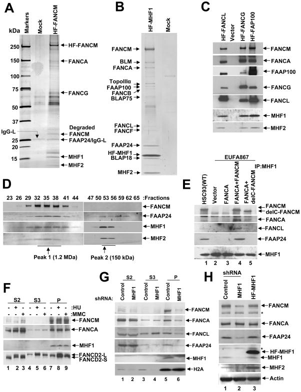
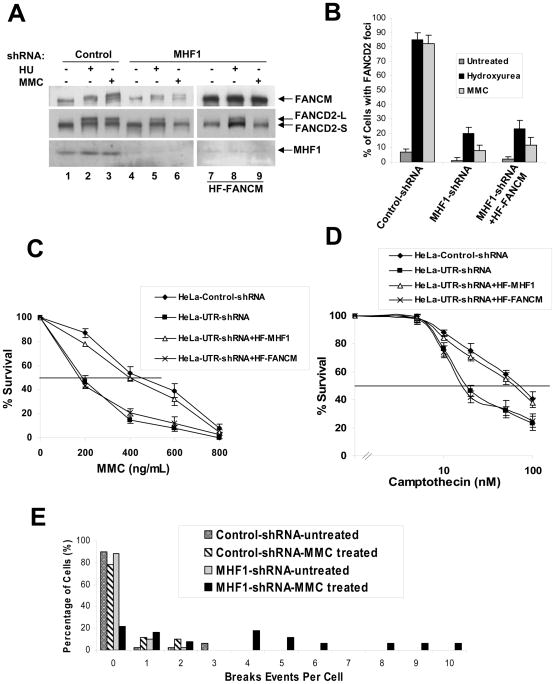
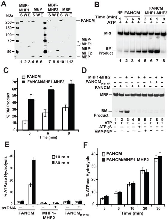
FANCM is a Fanconi anemia nuclear core complex protein required for the functional integrity of the FANC-BRCA pathway of DNA damage response and repair. Here we report the isolation and characterization of two histone-fold-containing FANCM-associated proteins, MHF1 and MHF2. We show that suppression of MHF1 expression results in (1) destabilization of FANCM and MHF2, (2) impairment of DNA damage-induced monoubiquitination and foci formation of FANCD2, (3) defective chromatin localization of FA nuclear core complex proteins, (4) elevated MMC-induced chromosome aberrations, and (5) sensitivity to MMC and camptothecin. We also provide biochemical evidence that MHF1 and MHF2 assemble into a heterodimer that binds DNA and enhances the DNA branch migration activity of FANCM. These findings reveal critical roles of the MHF1-MHF2 dimer in DNA damage repair and genome maintenance through FANCM.


Figures

Figure 1
Figure 1. MHF1 and MHF2 are components of the FA core complex
(A) Silver-stained gel showing the polypeptide bands purified from the nuclear extract of HT1080 cells transduced with vector alone (Mock) and cells expressing (His)6-FLAG-tagged (HF) FANCM. The polypeptides identified by MS analysis are indicated by an arrow. (B) A silver-stained gel showing the BRAFT complex components isolated from HeLa S3 cells expressing HF-MHF1. See also Fig. S1C and S1D. (C) Immunoblot showing the presence of MHF1 and MHF2 in immunoprecipitates of core complex proteins FANCL, FANCG and FAAP100. (D) Immunoblot showing co-fractionation of FAAP24, MHF1 and MHF2 with FANCM by Superose 6 gel filtration. (E) Immunoblot showing co-purification of FA core complex components with MHF1 from EUFA867 cells transduced with vector alone (lane 2), FANCA (lane 3), FANCA and FANCM (lane 4) and FANCA and delta (C-terminus deleted) FANCM (lane 5). (F) HeLa cells were treated with MMC (100 ng/mL for 16 hours) or HU (2 mM for 16 hours) and then fractionated into S2, S3, and P, as described in Material and Methods and immunoblotted with the indicated antibodies. (G) HeLa cells were transduced with control or MHF1 shRNAs, followed by cellular fractionation into S2, S3, and P fractions. The relevant proteins were revealed by immunoblotting. (H) HeLa cells were stably transduced with control-shRNA, MHF1-shRNA or HF-MHF1, and whole-cell lysates were analyzed by immunoblotting with the indicated antibodies. Asterisk (*) denotes a nonspecific cross-reactive band. See also Fig. S1.
Figure 2
Figure 2. MHF1 is required for the activation of the FA pathway
(A) Immunoblot showing that ablation of MHF1 expression in HeLa cells by shRNA reduces the level of monoubiquitinated FANCD2 in cells treated with MMC or HU. MHF1 knock down HeLa cells stably overexpressing HF-FANCM were used to show that phenotype observed is not due to the reduced levels of FANCM. (B) Histogram of immunofluorescence analysis of FANCD2 nuclear foci in HeLa cells in which MHF1 expression was knocked down by shRNA. Cells were either left untreated or exposed to MMC or HU, and the percentage of cells with 5 or more FANCD2 foci was determined by examining at least 150 cells. The result shows the average of 3 independent experiments with standard deviations. See also Fig. S2A. (C) & (D) Graph showing MMC and camptothecin survival curve of MHF1 knockdown cells. HeLa cells were transduced with either control or shRNA oligos targeting MHF1 and were subsequently treated with the indicated concentration of MMC and camptothecin. Visible colonies from 200 cells were counted after 10 days. The data represent the percent survival, as compared with untreated cells. Each experiment was performed in triplicate, and mean values are shown with standard deviations. HeLa cells stably expressing shRNA resistant HF-MHF1 were used as a control to show that the phenotype observed is not due to the off-target effect of shRNA. Also, MHF1 knock down HeLa cells stably overexpressing HF-FANCM were used to show that phenotype observed is not due to the reduced levels of FANCM. (E) Chromosomal aberrations following MMC treatment. HeLa cells were transfected with siRNAs and cells were treated with MMC. Metaphase spreads were prepared and scored for chromosomal aberrations. See also Fig. S2E.
Figure 3
Figure 3. DNA binding by the MHF1-MHF2 heterodimer
(A) Recombinant MHF1-(His)6-MHF2, MBP-MHF1, and MBP-MHF2 were expressed in and purified from E. coli. Purified proteins were analyzed by SDS-PAGE followed by Coomassie blue staining. (B) MHF1-(His)6-MHF2, MBP-MHF1 (1.0 and 2.0 μM), and MBP-MHF2 (1.0 and 2.0 μM) were examined for their DNA binding attribute with a mixture of the four 32P-labeled DNA substrates. (C). The indicated combinations of DNA substrates were incubated with MHF1-(His)6-MHF2 (50, 85, 120, 155, 190, 225, 270 and 305 nM). Quantification of the data is presented in the right panels. Note that treatment of nucleoprotein complexes with SDS and proteinase K (SDS/PK) released the DNA substrates. See also Fig. S3.
Figure 4
Figure 4. Enhancement of DNA branch migration by MHF1-MHF2 heterodimer
(A) Amylose beads pull-down assay with FANCM and MBP-MHF1 or MBP-MHF2. S, W, and E represent supernatant, wash and elution lanes. The asterisk (*) denotes a proteolytic product of FANCM. (B) Stimulation of the branch migration activity of FANCM by the MHF1-MHF2 dimer. ATP was present in all the reactions except the one in lane 8. NP - no protein control; BM, branch migration. (C) Quantification of the results shown in figure 4B. (D) DNA branch migration by FANCM, FANCMK117R, or the combination of FANCM or FANCMK117R and MHF1-MHF2 was examined with or without ATP, ATP-γ-S, or AMP-PNP, as indicated. BM, branch migration. (E) ATP hydrolysis by FANCM, MHF1-MHF2 or FANCMK117R was examined with or without ssDNA, as indicated. (F) Effect of MHF1-MHF2 on the ssDNA-dependent ATPase activity of FANCM. See also Fig. S3 and S4.

Comment in

References

    1. Ali AM, Kirby M, Jansen M, Lach FP, Schulte J, Singh TR, Batish SD, Auerbach AD, Williams DA, Meetei AR. Identification and characterization of mutations in FANCL gene: a second case of Fanconi anemia belonging to FA-L complementation group. Hum Mutat. 2009a;30:E761–770. - PMC - PubMed
    1. Ali AM, Singh TR, Meetei AR. FANCM-FAAP24 and FANCJ: FA proteins that metabolize DNA. Mutat Res. 2009b;668:20–26. - PMC - PubMed
    1. Amano M, Suzuki A, Hori T, Backer C, Okawa K, Cheeseman IM, Fukagawa T. The CENP-S complex is essential for the stable assembly of outer kinetochore structure. J Cell Biol. 2009;186:173–182. - PMC - PubMed
    1. Arents G, Moudrianakis EN. The histone fold: a ubiquitous architectural motif utilized in DNA compaction and protein dimerization. Proc Natl Acad Sci U S A. 1995;92:11170–11174. - PMC - PubMed
    1. Bakker ST, van de Vrugt HJ, Rooimans MA, Oostra AB, Steltenpool J, Delzenne-Goette E, van der Wal A, van der Valk M, Joenje H, Te Riele H, de Winter JP. Fancm-deficient mice reveal unique features of Fanconi anemia complementation group M. Hum Mol Genet 2009 - PubMed

Publication types