Review
. 2010 Mar;30(2):111-25.
doi: 10.1016/j.semnephrol.2010.01.003.

Genetics of focal segmental glomerulosclerosis and human immunodeficiency virus-associated collapsing glomerulopathy: the role of MYH9 genetic variation

Affiliations
Review

Genetics of focal segmental glomerulosclerosis and human immunodeficiency virus-associated collapsing glomerulopathy: the role of MYH9 genetic variation

Cheryl A Winkler et al. Semin Nephrol. 2010 Mar.

Abstract

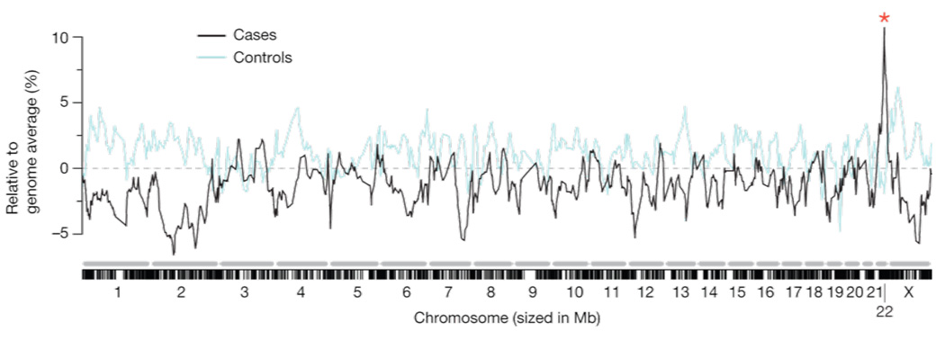
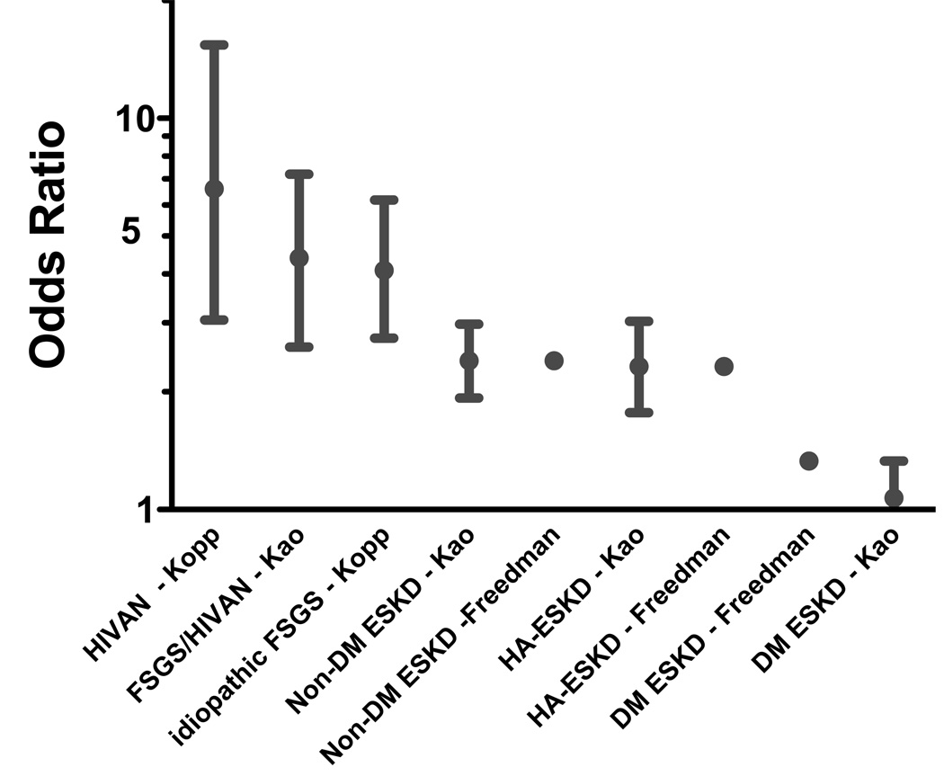
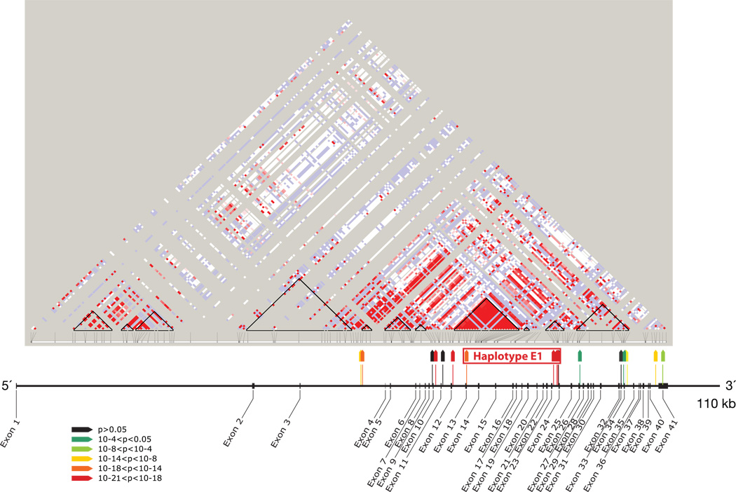
Until recently, knowledge of genetic causes of glomerular disease was limited to certain rare or uncommon inherited diseases, and to genes, either rare or with small effect, identified in candidate gene studies. These genetic factors accounted for only a very small fraction of kidney disease. However, the striking differences in frequency of many forms of kidney disease between African Americans and European Americans, which could not be explained completely by cultural or economic factors, pointed to a large unidentified genetic influence. Because focal segmental glomerulosclerosis (FSGS) and human immunodeficiency virus-associated collapsing glomerulopathy have striking racial disparities, we performed an admixture mapping study to identify contributing genetic factors. Admixture mapping identified genetic variants in the nonmuscle myosin heavy chain 9 gene (MYH9) as having a major influence on both FSGS and human immunodeficiency virus-associated collapsing glomerulopathy, with odds ratios from 4 to 8 and attributable fractions of 70% to 100%. Previously identified, rare, inherited MYH9 disorders point to a mechanism by which MYH9 variation disrupts the actin-myosin filaments responsible for maintaining the structure of podocytes, the cells that provide one of three filtration barriers in the glomeruli. MYH9 variation has a smaller but still highly significant effect on nondiabetic kidney disease, and a weaker but significant effect on diabetic kidney disease; it is unclear whether underlying cryptic FSGS is responsible for the MYH9 association with these diseases. The strong predicted power of MYH9 variation for disease indicates a clear role for genetic testing for these variants in personalized medicine, for assessment of genetic risk, and potentially for diagnosis.


Conflict of interest statement

Conflicts of interest. Three authors (CAW, GN, JBK) and the NIH have applied for a patent on SNPs associated with the diseases described here.

Figures

Figure 1
Figure 1. Admixture mapping for FSGS and HIVAN identifies a peak identifies a peak on chromosome 22
A summary of the genome-wide admixture scan for 412 FSGS and HIVAN cases and controls showing 92% African ancestry on chromosome 22 compared to a genome-wide average of 81%. The genome-wide lod score and case control lod scores were 9.2 and 12.4, respectively. Lod scores >2 are considered significant. Figure reproduced from Kopp et a. Nature Genetics 2008 with permission.
Figure 2
Figure 2. Actin-myosin and molecular partners in the podocyte
The actinomyosin complex is composed of an actin filament and a multimeric myosin assembly, composed of non-muscle myosin IIA or IIB molecules. The actinomyosin functional cycle is as follows: the myosin head binds ATP and this enables release from the actin filament, ATP is hydrolyzed to ADP and Pi, the myosin head domain binds actin when a site becomes available, the myosin head generates a power stroke that pushes the actin filament and simultaneously ADP and Pi are released, and myosin detaches from actin. This actinomyosin complex maintains cell tension and cytoarchitecture, and facilitates contraction (perhaps in response to increased hydrostatic glomerular capillary pressure) and migration (if podocyte foot processes are indeed mobile under physiologic or pathologic conditions). The figure shows links between the actinomyosin complex and four molecular complexes linking the actinomyosin complex to the extra-cellular environment: the slit diaphragm and podocalyxin (on the apical domain) and dystroglycan complex and a3b1 integrin (on the basal domain). The figure is not exhaustive in the molecular complexes nor in the inter-molecular interactions depicted. Molecular structures are depicted in varying levels of details and differing scales, and those interacting with nephrin are shown schematically. Further details on molecular interactions are available in an excellent review by Faul et al, Trends in Cell Biology 2007. Figure expanded from a previous figure, with permission (Kopp, Clin J Am Soc Nephrol 2009).
Figure 3
Figure 3
A comparison of odds ratios for HIVAN, idiopathic FSGS, non-diabetic ESKD, hypertension-attributed ESKD attributable, and diabetes-attributed ESKD in four published studies,,,.
Figure 4
Figure 4. MYH9 linkage disequilibrium and associations
Location and strength of association of 17 MYH9 SNPs, shown with map of exons of MYH9 and HapMap plot of MYH9 linkage disequilibrium (LD) in Yoruba from Nigeria. Strength of association for SNPs (colored wedges) is keyed by colors as shown. Black triangles on HapMap plot indicate HapMap-defined LD blocks. Haplotype E1 (for “extended haplotype 1”) extends through three HapMap LD blocks. F
Figure 5
Figure 5
Global distribution of MHY9 E1–5 extended haplotypes. Frequencies of risk (E1), protective (E2), and neutral MYH9 haplotypes (E3-E5) in the Human Genome Diversity Panel 1050 individuals in 51 populations, and grouped by the continental origins: African, European, Middle Eastern, South-Central Asian, East Asian, Oceanian, and American. African samples carry the largest proportion of risk haplotype, while most of the Europeans and the peoples in the Middle East in this study feature protective haplotype (E2). A large proportion of the South-Central Asian populations also have the protective haplotype, while the risk haplotype is at very low levels. East Asian and Oceanian populations are represented mainly by the neutral haplotype (E3), some protective haplotype, but with the risk haplotype virtually absent

Similar articles

Cited by

References

    1. DukeInstituteofGenomeSciencesandPolicy. Website Glossary. [Accessed 20 Sep 2009]. http://www.genomestohealth.org/glossary.php.
    1. Barisoni L, Schnaper HW, Kopp JB. Advances in the biology and genetics of the podocytopathies: implications for diagnosis and therapy. Arch Pathol Lab Med. 2009;133(2):201–216. - PMC - PubMed
    1. Woroniecki RP, Kopp JB. Genetics of focal segmental glomerulosclerosis. Pediatr Nephrol. 2007;22(5):638–644. - PMC - PubMed
    1. Bastian HM, et al. Systemic lupus erythematosus in three ethnic groups. XII. Risk factors fro lupus nephritis after diagnosis. lupus. 2002;11:152–160. - PubMed
    1. Burt VL, Whelton P, Roccella EJ, Brown C, Cutler JA, Higgins M, Horan MJ, Labarthe D. Prevalence of Hypertension in the US Adult Population : Results From the Third National Health and Nutrition Examination Survey, 1988–1991. Hypertension. 1995;25(3):305–313. - PubMed

Publication types

MeSH terms