

Review
doi: 10.1016/j.jamcollsurg.2010.01.020.
Clinical review of nonalcoholic steatohepatitis in liver surgery and transplantation
Affiliations
- PMID: 20347746
- PMCID: PMC4607044
- DOI: 10.1016/j.jamcollsurg.2010.01.020
Item in Clipboard
Review
Clinical review of nonalcoholic steatohepatitis in liver surgery and transplantation
J Am Coll Surg.
2010 Apr.
No abstract available
Figures

Histologic progression of nonalcoholic fatty liver disease from simple steatosis to cirrhosis.
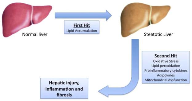
Progression of nonalcoholic fatty liver disease demonstrating the 2-hit hypothesis. The first hit consists of lipid accumulation resulting in simple benign steatosis. This primes the liver for a second hit, which can be multifactorial. This 2-hit process results in hepatic injury, inflammation, and ultimately fibrosis.

Histology of progression from steatosis to nonalcoholic steatohepatitis (NASH) cirrhosis. (A) Normal liver. Hematoxylin and eosin staining 200×. (B) Diffuse macrocytic steatosis with inflammatory changes. Hematoxylin and eosin staining 200×. (C) Macrocytic steatosis with neutrophil accumulation inflammation (NASH). Hematoxylin and eosin staining 400×. (D) NASH with pericellular fibrosis. Trichrome straining 400×. (E) NASH cirrhosis with bridging fibrosis. Trichrome staining 400×.
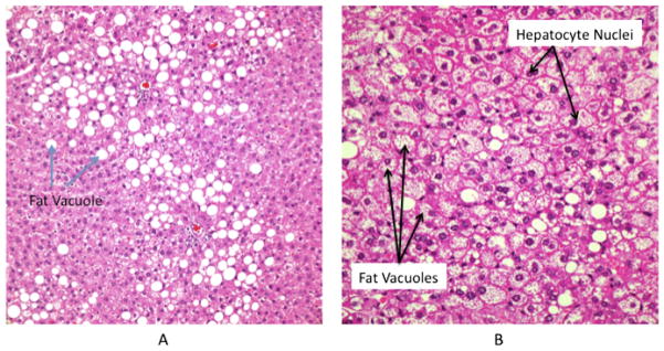
Histology of macrocytic and microcytic hepatic steatosis. (A) Macrocytic steatosis. Hematoxylin and eosin staining 400×. Fat vacuoles cause displacement of the hepatocyte nuclei. (B) Microcytic steatosis. Hematoxylin and eosin staining 400×. Small fat vacuoles clustered within hepatocyte with no displacement of nuclei.
Similar articles
-
Recurrent disease following liver transplantation for nonalcoholic steatohepatitis cirrhosis.Liver Transpl. 2009 Dec;15(12):1843-51. doi: 10.1002/lt.21943. Liver Transpl. 2009. PMID: 19938117
-
[Patomechanisms of hepatic steatosis].Orv Hetil. 2010 Feb 28;151(9):323-9. doi: 10.1556/OH.2010.28816. Orv Hetil. 2010. PMID: 20159747 Review. Hungarian.
-
[Nonalcoholic steatohepatitis: diagnosis, pathogenesis, treatment and prognosis].Ned Tijdschr Geneeskd. 2005 Feb 5;149(6):289-94. Ned Tijdschr Geneeskd. 2005. PMID: 15730035 Review. Dutch.
-
Long-term liver outcomes after metabolic surgery in compensated cirrhosis due to metabolic dysfunction-associated steatohepatitis.Nat Med. 2025 Mar;31(3):988-995. doi: 10.1038/s41591-024-03480-y. Epub 2025 Jan 27. Nat Med. 2025. PMID: 39870816
-
Non-alcoholic fatty liver disease.Bull Mem Acad R Med Belg. 2010;165(3-4):147-55; discussion 155-8. Bull Mem Acad R Med Belg. 2010. PMID: 21166266
Cited by
-
Inhibition of γ-glutamyltransferase ameliorates ischaemia-reoxygenation tissue damage in rats with hepatic steatosis.Br J Pharmacol. 2020 Nov;177(22):5195-5207. doi: 10.1111/bph.15258. Epub 2020 Oct 19. Br J Pharmacol. 2020. PMID: 32910829 Free PMC article.
-
Hepatic mitochondrial function analysis using needle liver biopsy samples.PLoS One. 2013 Oct 29;8(10):e79097. doi: 10.1371/journal.pone.0079097. eCollection 2013. PLoS One. 2013. PMID: 24205366 Free PMC article.
-
Strategies to rescue steatotic livers before transplantation in clinical and experimental studies.World J Gastroenterol. 2013 Aug 7;19(29):4638-50. doi: 10.3748/wjg.v19.i29.4638. World J Gastroenterol. 2013. PMID: 23922462 Free PMC article. Review.
-
Nonalcoholic fatty liver disease following liver transplantation.Hepatol Int. 2013 Jun;7(2):400-12. doi: 10.1007/s12072-013-9434-3. Epub 2013 Apr 26. Hepatol Int. 2013. PMID: 26201774
-
Phlorizin, an Important Glucoside: Research Progress on Its Biological Activity and Mechanism.Molecules. 2024 Feb 5;29(3):741. doi: 10.3390/molecules29030741. Molecules. 2024. PMID: 38338482 Free PMC article. Review.
References
-
- Angulo P. Nonalcoholic fatty liver disease. N Engl J Med. 2002;346:1221–1231. - PubMed
-
- Schaffner F, Thaler H. Nonalcoholic fatty liver disease [review] Prog Liver Dis. 1986;8:283–298. - PubMed
-
- Ludwig J, Viggiano TR, McGill DB, et al. Nonalcoholic steatohepatitis: Mayo Clinic experiences with a hitherto unnamed disease [see comment] Mayo Clin Proc. 1980;55:434–438. - PubMed
-
- Diehl AM, Goodman Z, Ishak KG. Alcohollike liver disease in nonalcoholics. A clinical and histologic comparison with alcohol-induced liver injury. Gastroenterology. 1988;95:1056–1062. - PubMed
-
- Brunt EM, Janney CG, Di Bisceglie AM, et al. Nonalcoholic steatohepatitis: a proposal for grading and staging the histological lesions. Am J Gastroenterol. 1999;94:2467–2474. - PubMed
Publication types
MeSH terms
Substances
Grants and funding
LinkOut - more resources
Full Text Sources
Medical
