. 2010 Jul;139(1):171-81.e9.
doi: 10.1053/j.gastro.2010.03.048. Epub 2010 Mar 27.

Esophageal squamous cell dysplasia and delayed differentiation with deletion of krüppel-like factor 4 in murine esophagus

Affiliations

Esophageal squamous cell dysplasia and delayed differentiation with deletion of krüppel-like factor 4 in murine esophagus

Marie-Pier Tetreault et al. Gastroenterology. 2010 Jul.

Abstract

Background & aims: Krüppel-like factor 4 (Klf; previously known a gut-enriched Krüppel-like factor) is a DNA-binding transcriptional regulator highly expressed in skin and gastrointestinal epithelia, specifically in regions of cellular differentiation. Homozygous null mice for Klf4 die shortly after birth from skin defects, precluding their analysis at later stages. The aim of this study was to analyze the function of Klf4 in keratinocyte biology and epithelial homeostasis in the adult by focusing on the squamous lined esophagus.

Methods: By using the ED-L2 promoter of Epstein-Barr virus to drive Cre, we obtained tissue-specific ablation of Klf4 in the squamous epithelia of the tongue, esophagus, and forestomach.

Results: Mice with loss of Klf4 in esophageal epithelia survived to adulthood, bypassing the early lethality. Tissue-specific Klf4 knockout mice had increased basal cell proliferation and a delay in cellular maturation; these mice developed epithelial hypertrophy and subsequent dysplasia by 6 months of age. Moreover, loss of Klf4 in vivo was associated with increased expression of the pro-proliferative Klf5, and Klf4 down-regulated Klf5 both transcriptionally and posttranscriptionally. By using gene expression profiling, we also showed decreased expression of critical late-stage differentiation factors and identified alterations of several genes important in cellular differentiation.

Conclusions: Klf4 is essential for squamous epithelial differentiation in vivo and interacts with Klf5 to maintain normal epithelial homeostasis.


Figures

Fig. 1
Fig. 1
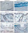
Klf4 was deleted successfully in esophageal epithelia of ED-L2/Cre;Klf4loxP/loxP mice. (A–B) Esophageal sections from Gt(ROSA)26tm1Sor control mice (A) showed no epithelial staining for β-galactosidase, while ED-L2/Cre;Gt(ROSA)26tm1Sor mice (B) had staining throughout the epithelia. (C–D) Both control (C) and ED-L2/Cre;Klf4loxP/loxP mice (D) demonstrated nuclear staining for Klf4 in the skin. (E–F) In esophageal epithelia, control mice (E) had nuclear Klf4 expression in cells of the suprabasal layer while Klf4 expression was lost in ED-L2/Cre;Klf4loxP/loxP mice (F). Scale bars (A–F), 10 μm.
Fig. 2
Fig. 2
ED-L2/Cre;Klf4loxP/loxP mutant mice had altered esophageal epithelial homeostasis. Compared to 1 month-old (A–B) and 3 month-old littermate controls (E–F), H&E stained esophageal epithelia from ED-L2/Cre;Klf4loxP/loxP mice were hypertrophic and keratinocytes showed altered morphology (C–D, G–H). The basal layers at these time points also appeared thickened and irregular. By 6 months of age, compared to littermate controls (I–J), ED-L2/Cre;Klf4loxP/loxP mice had evidence of delayed keratinocyte maturation and showed moderate dysplasia (K–L). Note the budding of epithelia into the lamina propria and the lack of surface keratinization in ED-L2/Cre;Klf4loxP/loxP mice. Scale bars (A, C, E, G, I, K), 50 μm; (B, D, F, H, J, L), 10 μm.
Fig. 3
Fig. 3
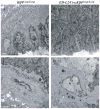
Ultrastructural abnormalities in Klf4-deficient esophageal epithelia from 3 month-old mice. Assessment of ultrastructure of littermate controls (A, C) and ED-L2/Cre;Klf4loxP/loxP mutant mice (B, D). (A–B) Compared to controls (A), esophageal epithelia of ED-L2/Cre;Klf4loxP/loxP mice (B) demonstrated changes in the orientation of cells as they progressed from the basal to suprabasal layers. (C–D) In the superficial layer, compared to controls (C), cells from ED-L2/Cre;Klf4loxP/loxP mice (D) had increased nuclear to cytoplasmic ratios. Typically, cells in this layer have small compacted nuclei. Scale bars (A–D), 2 μm.
Fig. 4
Fig. 4
ED-L2/Cre;Klf4loxP/loxP mice had increased proliferation and apoptosis in esophageal epithelia. (A–B) At 3 months of age, proliferating cells were confined to the basal layer in both control (A) and ED-L2/Cre;Klf4loxP/loxP mice (B). (C) Quantitation of BrdU-labeled cells revealed a statistically significant 1.9-fold increase in cell proliferation in ED-L2/Cre;Klf4loxP/loxP mice (p=0.006). (D–E) Compared to littermate controls (D), TUNEL staining of esophageal epithelia from ED-L2/Cre;Klf4loxP/loxP mice (E) demonstrated increased numbers of apoptotic cells in the superficial layers at 3 months of age. (F) By quantitation of TUNEL positive cells, apoptosis was increased 2.7-fold in ED-L2/Cre;Klf4loxP/loxP mice (p=0.001). Scale bars (A–B, D–E), 50 μm.
Fig. 5
Fig. 5
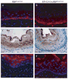
Klf4 deletion altered esophageal keratinocyte differentiation. (A–B) Staining for keratin 14 (red), which is normally confined to cells in the basal layer, as in controls (A), revealed expansion of the expression domain to include cells within the first few layers of suprabasal cells in ED-L2/Cre;Klf4loxP/loxP mice (B). (C–D) Compared to controls (C), expression of keratin 13, a marker of keratinocyte differentation, was decreased and shifted toward the luminal surface in ED-L2/Cre;Klf4loxP/loxP mice (D). (E–F) Keratin 4, another keratinocyte differentiation marker, was strongly expressed in the suprabasal layers of controls (E), but expression was markedly decreased in ED-L2/Cre;Klf4loxP/loxP mice (F). DAPI (blue) was used as a nuclear counterstain for immunofluorescence (A–B, E–F). Scale bars, 10 μm.
Fig. 6
Fig. 6
Klf4 inhibited expression of pro-proliferative Klf5. (A) Klf5 staining was seen in nuclei of cells only in the basal layer of the esophagus. (B) In contrast, ED-L2/Cre;Klf4loxP/loxP mice showed expression of Klf5 in both basal and suprabasal cells. (C) By qPCR, Klf5 expression was increased by 54% in esophageal epithelia of ED-L2/Cre;Klf4loxP/loxP mice (p=0.01). (D) ChIP assays of mouse primary esophageal keratinocytes demonstrated Klf4 binding to the 5′ regulatory region of Klf5 between −601 and −419. Cells were also treated with calcium to induce differentiation. Input was DNA extracted before immunoprecipitation. Anti-IgG antibody was used as a negative control. (E) Luciferase reporter assays with the 1 kb region immediately upstream of the Klf5 translational start site revealed a 53% decrease in activity (p<0.05) when cells were co-transfected with a Klf4 expression vector, compared to control. (F) Overexpression of Klf4 in mouse primary esophageal keratinocytes resulted in a 45% decrease in Klf5 mRNA by qPCR (p<1×10⁻⁹). (G) Western blotting and subsequent quantitation revealed a 98% decrease in Klf5 protein when Klf4 was overexpressed by 7-fold in these cells. A model for Klf4 and Klf5 in the switch from proliferation to differentiation in esophageal epithelia is shown in (H).

References

    1. Stappenbeck TS, Wong MH, Saam JR, et al. Notes from some crypt watchers: regulation of renewal in the mouse intestinal epithelium. Curr Opin Cell Biol. 1998;10:702–9. - PubMed
    1. Squier CA, Kremer MJ. Biology of oral mucosa and esophagus. J Natl Cancer Inst Monogr. 2001:7–15. - PubMed
    1. Seery JP. Stem cells of the oesophageal epithelium. J Cell Sci. 2002;115:1783–9. - PubMed
    1. Fuchs E, Horsley V. More than one way to skin. Genes Dev. 2008;22:976–85. - PMC - PubMed
    1. Katz JP, Kaestner KH. Cellular and molecular mechanisms of carcinogenesis. Gastroenterol Clin North Am. 2002;31:379–94. - PubMed

Publication types