. 2010 May 14;398(4):542-54.
doi: 10.1016/j.jmb.2010.03.039. Epub 2010 Mar 27.

Mapping of drebrin binding site on F-actin

Affiliations

Mapping of drebrin binding site on F-actin

Elena E Grintsevich et al. J Mol Biol. .

Abstract

Drebrin is a filament-binding protein involved in organizing the dendritic pool of actin. Previous in vivo studies identified the actin-binding domain of drebrin (DrABD), which causes the same rearrangements in the cytoskeleton as the full-length protein. Site-directed mutagenesis, electron microscopic reconstruction, and chemical cross-linking combined with mass spectrometry analysis were employed here to map the DrABD binding interface on actin filaments. DrABD could be simultaneously attached to two adjacent actin protomers using the combination of 2-iminothiolane (Traut's reagent) and MTS1 [1,1-methanediyl bis(methanethiosulfonate)]. Site-directed mutagenesis combined with chemical cross-linking revealed that residue 238 of DrABD is located within 5.4 A from C374 of actin protomer 1 and that native cysteine 308 of drebrin is near C374 of actin protomer 2. Mass spectrometry analysis revealed that a zero-length cross-linker, 1-ethyl-3-(3-dimethylaminopropyl) carbodiimide, can link the N-terminal G-S extension of the recombinant DrABD to E99 and/or E100 on actin. Efficient cross-linking of drebrin residues 238, 248, 252, 270, and 271 to actin residue 51 was achieved with reagents of different lengths (5.4-19 A). These results suggest that the "core" DrABD is centered on actin subdomain 2 and may adopt a folded conformation upon binding to F-actin. The results of electron microscopic reconstruction, which are in a good agreement with the cross-linking data, revealed polymorphism in DrABD binding to F-actin and suggested the existence of two binding sites. These results provide new structural insight into the previously observed competition between drebrin and several other F-actin-binding proteins.


Figures

Fig. 1
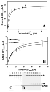
Fig. 1. Binding of the drebrin constructs to actin filaments
Binding affinity of drebrin constructs for F-actin was estimated by pelleting assays (see Materials and Methods). (a) Binding of the 1-300 drebrin construct to actin filaments (10 μM). Solid line corresponds to the best data fit. A Kd of the 1-300 drebrin construct for F-actin (0.17±0.005 μM) was calculated based on two independent experiments. (b) Binding affinities of N-GST fused DrABD constructs for F-actin (10 μM). Solid lines correspond to the best data fit with Kd = 6.6 ± 0.4 μM and 7.6 ± 0.6 μM for DrABD and DrABD300, respectively. Kd is an average value obtained in two independent experiments, as described in Materials and Methods. (c) Co-sedimentation of drebrin's ADF homology domain (drADF) with F-actin (Ac) (10 μM). (d) Control co-sedimentation of yeast cofilin (cof) with F-actin (10 μM) under the same conditions as in (d). Buffer composition: 5 mM MOPS, pH 7.2, 0.2 mM CaCl2, 0.4 mM EGTA, 0.2 mM ATP, 1 mM DTT, 50 mM KCl, 2 mM MgCl2. Lanes 1 – 6, 10 μM F-actin co-sedimented with 1.25, 2.5, 5, 10, 15, 25 μM of drebrin's ADF homology domain; lanes 9 – 14, 10 μM F-actin co-sedimented with 1.25, 2.5, 5, 10, 15, 25 μM of yeast cofilin; lanes 7 and 8, F-actin alone, supernatant and pellet, respectively.
Fig. 2
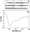
Fig. 2. Structure of DrABD
(a) Sequence of DrABD. Residues predicted to form helical structures are colored in gray (Jpred 3). The C-terminal extension that is truncated in DrABD300 construct is underlined. Two extra amino acids at N-terminus of the recombinant DrABD constructs are marked with asterisks. (b) CD spectrum (average of eight runs) of DrABD. Based on the results of two independent experiments, the secondary structure composition of DrABD is estimated to contain 28% helix, 15% β-sheets, 57% turns and random coil.
Fig. 3
Fig. 3. Electron microscopy and 3D-reconstruction of the drebrin-F-actin complex
Electron micrographs of F-actin alone (a) and (a*), filaments decorated with the DrABD construct (b) and drebrin 1-300 construct (b*). Three-dimensional reconstructions of pure F-actin (c) and five modes of binding of drebrin to F-actin (d - h). Atomic model of actin filament docked into each map is shown as blue ribbons (c - h). F-actin residues 99 and 100 (red), 51 (yellow), and 374 (green) are shown as spheres (c - h). An electron density envelope that corresponds to a globular protein containing 84 amino acid residues is shown as magenta meshwork (d - h). Comparison of the binding modes of DrABD and drebrin 1-300 construct to F-actin (d*) – (h*). Reconstructions of F-actin decorated with DrABD are shown as transparent surfaces, while volumes resulted from filaments complexed with the 1-300 construct containing both DrABD and AFD-homology domain are shown as blue meshwork. Atomic model of actin filament docked into each map is shown as blue ribbons (d* - h*). F-actin residues 99 and 100 (red), 51 (yellow), and 374 (green) are shown as spheres (a - e). An electron density envelope that corresponds to a globular protein comprised of 84 amino acid residues is shown as magenta solid surface. The modes of binding obtained for drebrin 1-300 construct are similar to the ones observed for isolated DrABD
Fig. 4
Fig. 4. DrABD is in close proximity to the C-terminal regions of two adjacent actin protomers
(a) DrABD treated with 2-iminothiolane (Traut's reagent) can be covalently attached to MTS1 pre-modified skeletal F-actin: lane 1 – MTS1 modified FA (10 μM) (A), lane 2 - 2-iminothiolane-treated DrABD (20 μM) incubated for 5 min. with MTS1 modified actin. Two main cross-linking products were detected: actin-DrABD heterodimer (AD) and the species that according to molecular weight and mass spectrometry analysis corresponds to two actin protomers and one DrABD (ADA). (b) Native C308 of DrABD is within 5.4 Å from C374 on actin: lanes 1 – 3 - skeletal F-actin; lanes 4 - 6 - WT yeast actin; lanes 7 – 9 - yeast actin mutant C374A. Lanes 1, 4, 7 - MTS1 modified actins; lanes 2, 5, 8 - MTS1 modified actins in the presence of DrABD (5 min reaction time); lanes 3, 6, 9 – same as lanes 2, 5, 8 but reaction time is 17 min. The final concentrations of actin and DrABD were 9.5 μM and 28.5 μM, respectively.
Fig. 5
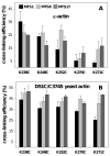
Fig. 5. Mapping DrABD binding interface on F-actin
(A) Thiol specific cross-linking of five DrABD mutants to skeletal F-actin (a) or to yeast actin mutant D51C/C374S (b) modified with MTS reagents of different length. Drebrin's residue 238 is within 5.4 Å from the Cys 374 of actin (C-terminus). Drebrin's residues 238, 248, 252, 270 and 271 are within ∼12.1 Å from residue 51 on actin (D-loop, subdomain 2). F-actin was pre-modified with MTS immediately prior to the cross-linking. The final concentrations of actin and DrABD were 10 μM and 30 μM, respectively. The reactions were stopped with NEM after 5 min and the resulting mixtures were analyzed by SDS PAGE. Relative intensities of protein bands were determined by densitometric analysis. Cross-linking efficiencies were estimated as follows: [actin total (before the reaction) – uncross-linked actin monomer left after 5 min]/total actin, %. Black: MTS1 (5.4 Å); light grey: MTS8 (12.1 Å); dark grey: MTS17 (19 Å).
Fig. 6
Fig. 6. N-terminus of DrABD constructs can be attached to Subdomain 1 of actin with zero length cross-linking reagent, EDC
(a) Tryptic peptides of DrABD-actin hetorodimer were analyzed by tandem mass spectrometry. The 663.61 (M +4H)4+ peak in MS spectra, corresponding to cross-linked peptides, was fragmented using ESI-MS/MS (Waters Synapt QTOF mass spectrometer). Some of the identified fragments are indicated in the figure. The first digit indicates the peptide from which the fragment originates (1 = actin and 2 = drebrin), the letter refers to the ion series and the last digit to the ion number. Hyphenated labels indicate cross-linked fragments with the left fragment originating from actin and the right one from drebrin. M represents intact drebrin peptide. The inset shows a schematic representation of the cross-linked peptides. In the scheme, the cross-linked residues are connected with a solid line (major site), a minor cross-linking site on actin peptide is marked with the asterisk. (b) Internal ions of the cross-linked peptides support the attachment of N-terminal Gly of DrABD to E99/E100 (major population) and E107 (minor cross-link) on actin.
Fig. 7
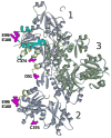
Fig. 7. Summary of the cross-linking results
Holmes model of actin filament structure (36). Actin protomers are marked as 1, 2 and 3. Actin residues involved in cross-linking with DrABD are shown in magenta. Actin peptides involved in interactions with cofilin (17) and α-actinin (32) are shown in yellow and blue (Protomer 1), respectively. The overlap between cofilin and α-actinin binding sites is shown in green (Protomer 1)
Fig. 8
Fig. 8. Single cysteine substitutions introduced into DrABD do not impair its ability to bind F-actin
Time course of cross-linking reactions (0, 5, 10, 20, 40 and 75 min) between skeletal F-actin (10μM) and DrABD300 mutants (30μM) in the presence of EDC (30μM). The higher mobility bands correspond to actin; the lower mobility bands represent the cross-linked actin-DrABD complex. Reaction conditions: 5 mM MOPS pH 7.2, 0.2 mM CaCl2, 0.2 mM ATP, 0.11 mM TCEP, 100 mM KCl, 2 mM MgCl2.
Scheme 1
Scheme 1

Similar articles

Cited by

References

    1. Sekino Y, Kojima N, Shirao T. Role of actin cytoskeleton in dendritic spine morphogenesis. Neurochem Int. 2007;51:92–104. - PubMed
    1. Kojima N, Shirao T. Synaptic dysfunction and disruption of postsynaptic drebrin-actin complex: A study of neurological disorders accompanied by cognitive deficits. Neurosci Res. 2007;58:1–5. - PubMed
    1. Peitsch WK, Hofmann I, Pratzel S, Grund C, Kuhn C, Moll I, Langbein L, Franke WW. Drebrin particles: components in the ensemble of proteins regulating actin dynamics of lamellipodia and filopodia. Eur J Cell Biol. 2001;80:567–579. - PubMed
    1. Peitsch WK, Hofmann I, Endlich N, Pratzel S, Kuhn C, Spring H, Grone HJ, Kriz W, Franke WW. Cell biological and biochemical characterization of drebrin complexes in mesangial cells and podocytes of renal glomeruli. J Am Soc Nephrol. 2003;14:1452–1463. - PubMed
    1. Grenklo S, Hillberg L, Zhao Rathje LS, Pinaev G, Schutt C, Lindberg U. Tropomyosin assembly intermediates in the control of microfilament system turnover. Eur J Cell Biol. 2008;87:905–920. - PubMed

Publication types