. 2010 Jul;47(1):65-73.
doi: 10.1016/j.bone.2010.03.012. Epub 2010 Mar 27.

Bone morphogenetic protein 2 stimulates endochondral ossification by regulating periosteal cell fate during bone repair

Affiliations

Bone morphogenetic protein 2 stimulates endochondral ossification by regulating periosteal cell fate during bone repair

Yan Yiu Yu et al. Bone. 2010 Jul.

Abstract

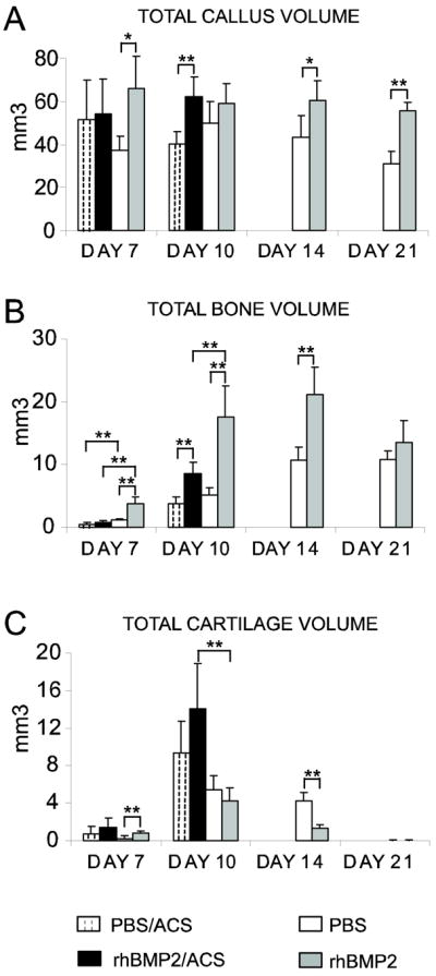
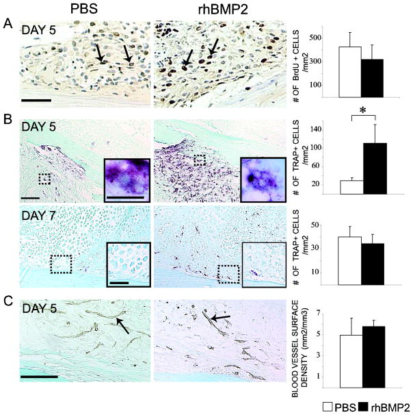
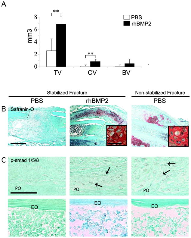
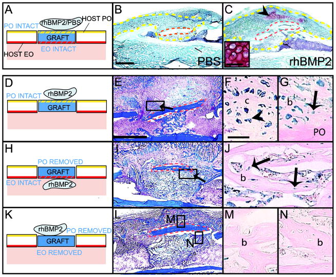
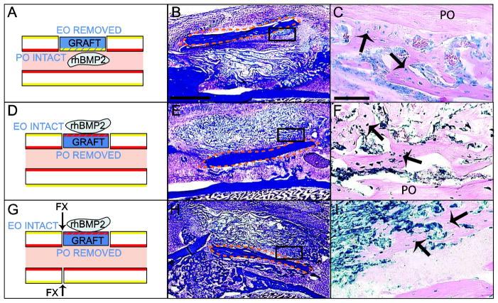
Bone repair depends on the coordinated action of numerous growth factors and cytokines to stimulate new skeletal tissue formation. Among all the growth factors involved in bone repair, Bone Morphogenetic Proteins (BMPs) are the only molecules now used therapeutically to enhance healing. Although BMPs are known as strong bone inducers, their role in initiating skeletal repair is not entirely elucidated. The aim of this study was to define the role of BMP2 during the early stages of bone regeneration and more specifically in regulating the fate of skeletal progenitors. During healing of non-stabilized fractures via endochondral ossification, exogenous BMP2 increased the deposition and resorption of cartilage and bone, which was correlated with a stimulation of osteoclastogenesis but not angiogenesis in the early phase of repair. During healing of stabilized fractures, which normally occurs via intramembranous ossification, exogenous BMP2 induced cartilage formation suggesting a role in regulating cell fate decisions. Specifically, the periosteum was found to be a target of exogenous BMP2 as shown by activation of the BMP pathway in this tissue. Using cell lineage analyses, we further show that BMP2 can direct cell differentiation towards the chondrogenic lineage within the periosteum but not the endosteum, indicating that skeletal progenitors within periosteum and endosteum respond differently to BMP signals. In conclusion, BMP2 plays an important role in the early stages of repair by recruiting local sources of skeletal progenitors within periosteum and endosteum and by determining their differentiation towards the chondrogenic and osteogenic lineages.


Figures

FIG. 1
FIG. 1
Effects of rhBMP2 treatment during the course of non-stabilized tibial fracture repair. Histomorphometric analyses of total callus volume (TV, A), total bone volume (BV, B), and total cartilage volume (CV, C in PBS/ACS, rhBMP2/ACS, PBS, and rhBMP2-treated mice at days 7, 10, 14 and 21 post-fracture (n=5 per group). *p< 0.05, **p<0.01. Bars represent mean ± s.d.
FIG. 2
FIG. 2
Effects of rhBMP2 on cell proliferation, osteoclastogenesis and angiogenesis during non-stabilized fracture repair. (A) BrdU immunohistochemistry staining (left, arrows) and stereological analyses of BrdU-positive cells per area of fracture callus (right, n=5 per group) in PBS- and rhBMP2-treated calluses at day 5 post-fracture. (B) TRAP staining (left) and stereology analyses of TRAP-positive cells per area of fracture callus (right, n=5 per group) in PBS- and rhBMP2-treated callus at days 5 and 7 post-fracture. There is a significantly increase in the number of TRAP-positive cells in rhBMP2-treated calluses compared to controls at day 5 (*p<0.05). Inserted boxes show high magnifications of multinucleated TRAP-positive cells in the bone marrow of rhBMP2- and PBS-treated calluses (B, top) and hypertrophic cartilage association with TRAP-positive cells in the periosteum of rhBMP2-treated samples (B, bottom). (C) PECAM immunohistochemistry (left, arrows) and stereology analyses of the surface density of blood vessel within the callus (right, n=5 per group) in PBS- and rhBMP2-treated calluses at day 5 post-fracture. Scale bars: (A) = 50μm; (B) = 100 μm; (B, inserted box, top) = 10μm; (B, inserted box, bottom) = 50μm; (C) = 100μm.
FIG. 3
FIG. 3
rhBMP2 induces cartilage formation during stabilized fracture repair and activates BMP signaling pathway within periosteum. (A) Histomorphometric measurements of total callus (TV), total cartilage volume (CV) and total bone volume (BV) in PBS- and rhBMP2-treated calluses. There is a statistically significant increase in total callus volume and cartilage volume in rhBMP2 treated calluses compared with PBS-treated calluses (**p<0.01). (B) Safranin-O/Fast Green (SO) staining of sections through the callus of (left) PBS- and (middle) rhBMP2-treated stabilized fractures and (right) PBS-treated non-stabilized fractures at day 10 post-fracture. Inserted boxes show high magnification of chondrocytes. (C) Negative p-Smad 1/5/8 immunostaining in the periosteum (PO, top) of stabilized fractures treated with PBS (left) and positive staining in cells of the activated periosteum in rhBMP2- treated stabilized fractures (middle, arrows) and PBS-treated non-stabilized fractures (right, arrows) at day 5. Negative p-Smad 1/5/8 immunostaining in endosteum (EO, bottom). Scale bars (B) = 1mm; (B, inserted boxes) and (C) = 50 μm,
FIG. 4
FIG. 4
Distinct effects of rhBMP2 on periosteum- and endosteum-derived cells during bone repair. (A) Schematic representation of Rosa 26 bone grafts (blue) treated with PBS/ACS or rhBMP2/ACS and transplanted in the cortex of wild type host tibia. Safranin-O/Fast green (SO) staining at day 10 of (B) PBS/ACS and (C) rhBMP2/ACS treated samples. Cartilage (arrowhead and high magnification) is found at the periosteal surface of rhBMP2/ACS-treated samples (C). (D, H, K) (Left) Schematic representations of Rosa 26 bone grafts (blue) treated with rhBMP2/ACS transplanted in the cortex of wild type host tibia. (Middle) Longitudinal sections through the bone graft stained with Trichrome (TC) and (Right) adjacent sections stained with X-gal at 10 days following bone grafting. (F–G) X-gal-positive osteoblasts and osteocytes (arrow) and pre-hypertrophic chondrocytes (arrowhead) are found at the intact periosteal surface in rhBMP2-treated samples. (J) X-gal-positive osteoblasts and osteocytes (arrow) are found at the intact endosteal surface within bone marrow cavity but (M–N) not in negative controls. Dotted yellow lines delimit the callus. Dotted orange lines delimit the graft. c= cartilage; b=bone. Scale bars (B, C, E, I, L) = 1mm; (C, inserted box) = 50μm; (F, G, J, M and N) = 100μm.
FIG. 5
FIG. 5
Effect of the tissue environment on periosteal and endosteal responses to rhBMP2 during bone repair. (Left) Schematic representation of Rosa26 bone grafts (blue) treated with rhBMP2/ACS. Intact periosteum (PO, yellow) was placed in the environment of bone marrow (A) and intact endosteum (EO, red) was placed in the environment of periosteum (D, G). Arrows indicate where the fracture is created (G). (Middle) Sections through the bone graft were stained with Trichrome (TC) and (Right) adjacent sections were stained with X-gal at 10 days following bone grafting. (A–C) During bone graft healing, X-gal-positive osteoblasts and osteocytes (arrows) derived from PO are found at the periosteal surface of the graft in the host bone marrow. (D–F) During bone graft and (G–I) non-stabilized fracture healing, X-gal-positive osteoblasts and osteocytes (arrows) derived from EO are located at the endosteal surface of the graft in the environment of the host PO. Dotted orange lines delimit the bone graft. Scale bars (B, E, H) = 1mm; (C, F, I) = 100μm.

Similar articles

Cited by

References

    1. Hogan BL. Bone morphogenetic proteins: multifunctional regulators of vertebrate development. Genes Dev. 1996;a(13):1580–94. - PubMed
    1. Huang YH, et al. Bone morphogenetic proteins and osseointegration: current knowledge - future possibilities. Periodontol 2000. 2008;47:206–23. - PubMed
    1. Linkhart TA, Mohan S, Baylink DJ. Growth factors for bone growth and repair: IGF, TGF beta and BMP. Bone. 1996;19(1 Suppl):1S–12S. - PubMed
    1. Kugimiya F, Kawaguchi H, Chung UI. BMP and bone formation. Clin Calcium. 2004;14(1):173–9. - PubMed
    1. Sellers RS, et al. Repair of articular cartilage defects one year after treatment with recombinant human bone morphogenetic protein-2 (rhBMP-2) J Bone Joint Surg Am. 2000;82(2):151–60. - PubMed

Publication types

MeSH terms

Substances