. 2010 Jun 11;285(24):18155-65.
doi: 10.1074/jbc.M109.064931. Epub 2010 Mar 26.

Oxidative activity of yeast Ero1p on protein disulfide isomerase and related oxidoreductases of the endoplasmic reticulum

Affiliations

Oxidative activity of yeast Ero1p on protein disulfide isomerase and related oxidoreductases of the endoplasmic reticulum

Elvira Vitu et al. J Biol Chem. .

Abstract

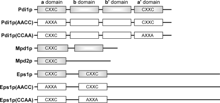
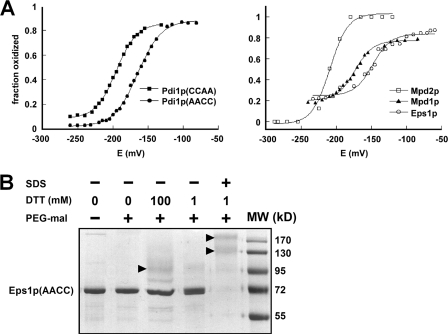
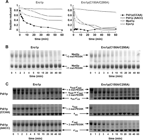
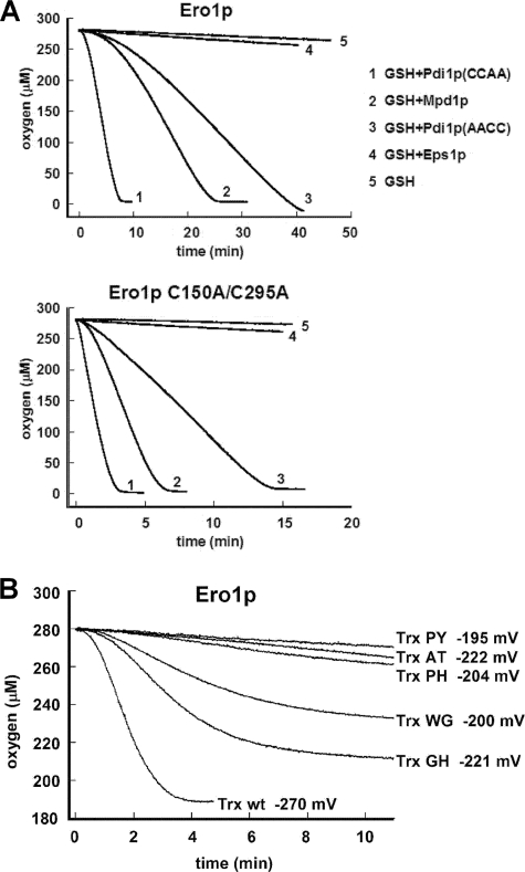
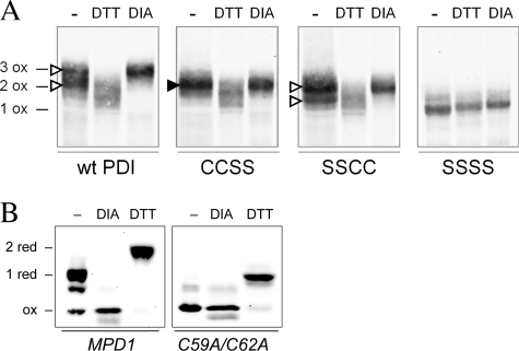
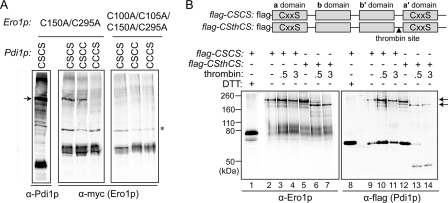
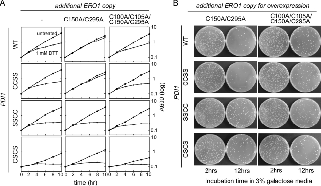
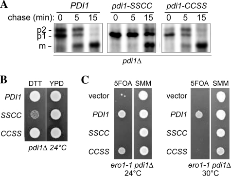
The sulfhydryl oxidase Ero1 oxidizes protein disulfide isomerase (PDI), which in turn catalyzes disulfide formation in proteins folding in the endoplasmic reticulum (ER). The extent to which other members of the PDI family are oxidized by Ero1 and thus contribute to net disulfide formation in the ER has been an open question. The yeast ER contains four PDI family proteins with at least one potential redox-active cysteine pair. We monitored the direct oxidation of each redox-active site in these proteins by yeast Ero1p in vitro. In this study, we found that the Pdi1p amino-terminal domain was oxidized most rapidly compared with the other oxidoreductase active sites tested, including the Pdi1p carboxyl-terminal domain. This observation is consistent with experiments conducted in yeast cells. In particular, the amino-terminal domain of Pdi1p preferentially formed mixed disulfides with Ero1p in vivo, and we observed synthetic lethality between a temperature-sensitive Ero1p variant and mutant Pdi1p lacking the amino-terminal active-site disulfide. Thus, the amino-terminal domain of yeast Pdi1p is on a preferred pathway for oxidizing the ER thiol pool. Overall, our results provide a rank order for the tendency of yeast ER oxidoreductases to acquire disulfides from Ero1p.


Figures

FIGURE 1.
FIGURE 1.
Domain organization of yeast PDI family proteins and location of Cys-X-X-Cys motifs. Standard nomenclature for Pdi1p domains (a, b, b′, and a′ domains) is indicated. Boxes correspond to trx domains. Gray boxes distinguish wild-type proteins from mutants used in this study, which are shown with white boxes.
FIGURE 2.
FIGURE 2.
Redox characteristics of yeast PDI family Cys-X-X-Cys motifs. A, redox potential of the samples was adjusted by varying the GSH/GSSG ratio at pH 7.0. After equilibration in redox buffer, the proteins were precipitated and reacted with mal-PEG5K, separated by SDS-PAGE, and visualized using fluorescent stain. The intensities of the bands were quantified and plotted as fraction oxidized. Data for the five Cys-X-X-Cys motifs are shown on two graphs for clarity. Each titration was fitted to the Nernst equation for a two-electron transfer to give reduction potential values of Pdi1p(CCAA): −194 mV, Pdi1p(AACC): −164 mV, Mpd1p: −174 mV, and the Eps1p amino-terminal domain: −149 mV. The Mpd2p reduction potential is estimated to be −210 mV. B, the Cys-X-X-Cys of the second Eps1p trx domain is not fully reduced by high concentrations of DTT in the folded state. Alkylation of free thiols with mal-PEG5K was used to distinguish oxidized from reduced fractions after the indicated treatments. The Eps1p(AACC) mutant was incubated with low (1 mm) or high (100 mm) DTT concentrations in the folded state or with 1 mm DTT after denaturing the protein with detergent. In the folded state, the protein was highly resistant to reduction. Only in the denatured state did substantial reduction occur, as indicated by the disappearance of the unmodified protein (labeled Eps1p(AACC) to the side of the gel) and the appearance of species of higher apparent molecular weights (indicated by arrowheads). Eps1p(AACC) contains six cysteines, so the various higher molecular weight species may have different subsets of these cysteines reduced and modified by mal-PEG5K.
FIGURE 3.
FIGURE 3.
Direct oxidation rates of PDI family proteins by Ero1p monitored by gel assays. A, reduced PDI family proteins (150 μm thiols) were mixed with 2 μm Ero1p or Ero1p(C150A/C295A). Loss of reactivity with mal-PEG5K upon disulfide bond formation was monitored by quantification of the accumulating unmodified protein after separation by SDS-PAGE. Lag phases are less evident in this experiment than in Fig. 4, because of the poor time resolution of gel-based assays. Furthermore, due to the relatively low substrate/enzyme ratio in this experiment (37.5:1), a significant fraction (estimated 5–10%) of substrate is oxidized by dithiol/disulfide exchange with the Ero1p shuttle and regulatory disulfides prior to oxygen consumption and enzyme turnover. B, except for a lower Mpd2p concentration, the experiment was conducted as in A, but the gels, rather than results of quantification, are shown. Mpd2p was partially reduced by incubation with 23 mm GSH for 1 h. Following removal of GSH, Mpd2p (50 μm thiols) was mixed with 2 μm Ero1p or Ero1p(C150A/C295A). The Mpd2p protein lacking its signal sequence has seven cysteine residues. One unpaired cysteine reacts with mal-PEG5K regardless of the redox status of the Cys-X-X-Cys motif. C, the two Pdi1p active sites appear to function independently during oxidation by Ero1p, because the two oxidation events seen for wild-type Pdi1p (top panel) occur at similar rates as the oxidation of the Pdi1p(CCAA) (middle panel) and Pdi1p(AACC) (bottom panel) mutants. This observation holds for both wild-type Ero1p and the C150A/C295A mutant. Bands are labeled according to the state of each Cys-X-X-Cys motif present (subscript “red” indicates reduced and “ox” oxidized) and the number of mal-PEG5K additions present. The gels shown here, which are similar to those used to obtain the data in A, further demonstrate that only a single disulfide was reduced initially in Pdi1p(AACC) and Pdi1p(CCAA), and two disulfides in wild-type Pdi1p, indicating that the Cys-X6-Cys motif remained oxidized as intended.
FIGURE 4.
FIGURE 4.
Oxidation rates measured by oxygen consumption. A, reduced Pdi1p(CCAA), Pdi1p(AACC), Mpd1p, or Eps1p at an initial thiol concentration of 100 μm were mixed with 10 mm GSH. At time zero, Ero1p or Ero1p(C150A/C295A) was added to a concentration of 1 μm. Representative progress curves are shown, but in some experiments, oxidation of Mpd1p was slightly slower. Lag phases are evident in this experiment. B, oxidation of E. coli thioredoxin and mutant series. Reduced thioredoxin or active-site mutants (the two letters indicate the identity of the X-X amino acids in the Cys-X-X-Cys motif) at 200 μm thiol concentration were oxidized by 1 μm Ero1p.
FIGURE 5.
FIGURE 5.
Redox states of Pdi1p and Mpd1p in vivo. A, wild-type Pdi1p and mutants Pdi1p(CCSS), Pdi1p(SSCC), and Pdi1p(SSSS) were purified from yeast cell lysates that had been reacted with NEM to block free thiols. Purified proteins were subsequently reduced with DTT and modified by mal-PEG2K. Pdi1p was visualized by Western blotting with anti-Pdi1p serum. With this protocol, the presence of a disulfide bond in the original cell lysate is reflected by mal-PEG2K modification and a corresponding decreased migration during SDS-PAGE. Fully oxidized Pdi1p controls (e.g. three disulfide bonds for wild-type Pdi1p (3 ox)) were prepared by lysis of cells in the presence of 10 mm diamide (DIA). Reduced migration controls were prepared by cell lysis in the presence of DTT; note that these lysis conditions did not result in reduction of the structural disulfide in Pdi1p as evident by the co-migration of the DTT and diamide-treated samples for Pdi1p(SSSS). The filled arrowhead indicates the single band representing a species with an oxidized a domain in the mutant lacking the active-site cysteines of the a′ domain. The open arrowheads indicate the two bands corresponding to the oxidized and reduced forms of the a′ domain in the mutant lacking the active-site cysteines of the a domain. B, yeast mpd1Δ cells containing plasmids encoding Myc-tagged Mpd1p or an Mpd1p active-site mutant, Mpd1p(C59A/C62A), were grown in SMM at 30 °C. The oxidation state of Mpd1p was assessed after resolving mal-PEG2K-modified cellular proteins by reducing SDS-PAGE and immunoblotting with anti-Myc. Using this protocol, free cysteine thiols within Mpd1p in the original cell lysate result in mal-PEG2K modification and a corresponding decrease in migration. Oxidized and reduced controls were prepared by treatment of cell lysates with diamide or DTT prior to mal-PEG2K.
FIGURE 6.
FIGURE 6.
Ero1p preferentially forms a mixed-disulfide intermediate with the a domain of Pdi1p. A, formation of mixed-disulfide intermediates between Ero1p(C150A/C295A) (with a Myc epitope tag) and Pdi1p mutants Pdi1p(CSCS), Pdi1p(CSCC), and Pdi1p(CCCS). NEM-modified yeast cell lysates were resolved by non-reducing SDS-PAGE, and mixed-disulfide intermediates between Ero1p and Pdi1p were identified by Western blotting with either anti-Pdi1p or anti-Myc. Catalytically inactive Ero1p(C100A/C105A/C150A/C295A) was used as a negative control. An arrow denotes the mixed-disulfide complexes between mutant Ero1p and a subset of Pdi1p variants. Other bands in the leftmost-Pdi1p) lane are mixed-disulfide complexes between Pdi1p(CSCS) and other proteins (high molecular weight bands) or free Pdi1p(CSCS) (intense lower band). The asterisk indicates a nonspecific background band that does not disappear upon reduction (not shown). B, thrombin digestion of the Pdi1p(CSCS)-Ero1p(C150A/C295A) mixed-disulfide intermediate. To enable dissection of Pdi1p(CSCS), a thrombin site was introduced into the segment linking the b′ and a′ domains to generate the construct Pdi1p(CSthCS). Mixed-disulfide intermediates were isolated under non-reducing conditions from cells expressing a FLAG-epitope-tagged Pdi1p by affinity to anti-FLAG beads. Purified Pdi1p(CSCS)- and Pdi1p(CSthCS)-Ero1p(C150A/C295A) mixed-disulfide complexes were digested by thrombin, and association of the Pdi1p fragments with Ero1p was analyzed by immunoblotting with either anti-Ero1p or anti-FLAG. Arrows highlight the major Pdi1p-Ero1p mixed-disulfide complexes observed before (top) and after (bottom) thrombin cleavage.
FIGURE 7.
FIGURE 7.
Disulfide relay system between Ero1p and the a domain of Pdi1p. A, the de-regulated Ero1p(C150A/C295A) mutant requires a functional Pdi1p a domain to suppress growth defects observed upon addition of 1 mm DTT. Yeast strains CKY1046–1049 were transformed with Ero1p(C150A/C295A) (pCS456) or the catalytically inactive Ero1p(C100A/C105A/C150A/C295A) (pCS483). Yeast strains were cultured in SMM at 30 °C, and growth was followed by light scattering (at 600 nm). At 2 h, DTT was added to half the cultures to a final concentration of 1 mm. B, cellular toxicity by overexpressed Ero1p(C150A/C295A) is alleviated by disruption of the Pdi1p a domain. CKY1046–1049 were transformed with a plasmid encoding Ero1p(C150A/C295A) (pCS452) or catalytically inactive Ero1p(C100A/C105A/C150A/C295A) (pCS504) under the control of the GAL promoter. After 2 and 12 h of growth in SMM with 3% galactose, 10−4 A600 unit of cells were spread onto SMM plates and incubated for 2 days at 30 °C. Cell viability was calculated by comparing colony forming units between cells expressing Ero1p(C150A/C295A) and cells expressing Ero1p(C100A/C105A/C150A/C295A) 12 h after induction.
FIGURE 8.
FIGURE 8.
Pdi1p a domain mutant shows diminished oxidative folding capacity. A, processing of CPY in pdi1Δ cells containing PDI1 plasmids. Cells were pulse labeled for 7 min and chased at 30 °C, and CPY was immunoprecipitated and resolved by SDS-PAGE. ER (p1), Golgi (p2), and vacuolar (m) forms of CPY are indicated. B, yeast strains from panel A were grown for 12 h in SMM without leucine and spotted onto YPD plates with or without 10 mm DTT. Strains were grown for 2 days at 24 °C. C, the pdi1Δ strain CKY1058 covered by an URA3-marked PDI1 plasmid was transformed with LEU2-marked plasmids encoding Pdi1p (pCS463), Pdi1p(SSCC) (pCS465), Pdi1p(CCSS) (pCS464), or empty vector. These strains were spotted onto SMM plates or SMM plates containing 5-fluoroorotic acid (to select for yeast able to grow in the absence of the URA3 plasmid). Plates were incubated at 24 °C, 30 °C, or 37 °C for 3 days. All four strains were inviable at 37 °C (not shown), consistent with the previously characterized lethality of the ero1-1 mutant at 37 °C (1).

Similar articles

Cited by

References

    1. Frand A. R., Kaiser C. A. (1998) Mol. Cell 1, 161–170 - PubMed
    1. Pollard M. G., Travers K. J., Weissman J. S. (1998) Mol. Cell 1, 171–182 - PubMed
    1. Frand A. R., Kaiser C. A. (1999) Mol. Cell 4, 469–477 - PubMed
    1. Jessop C. E., Bulleid N. J. (2004) J. Biol. Chem. 279, 55341–55347 - PubMed
    1. Molteni S. N., Fassio A., Ciriolo M. R., Filomeni G., Pasqualetto E., Fagioli C., Sitia R. (2004) J. Biol. Chem. 279, 32667–32673 - PubMed

Publication types

LinkOut - more resources