

The presence of a single N-terminal histidine residue enhances the fusogenic properties of a Membranotropic peptide derived from herpes simplex virus type 1 glycoprotein H
- PMID: 20348105
- PMCID: PMC2878091
- DOI: 10.1074/jbc.M110.114819
The presence of a single N-terminal histidine residue enhances the fusogenic properties of a Membranotropic peptide derived from herpes simplex virus type 1 glycoprotein H
Abstract
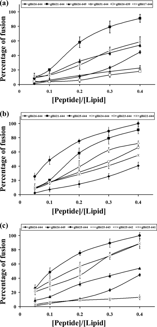
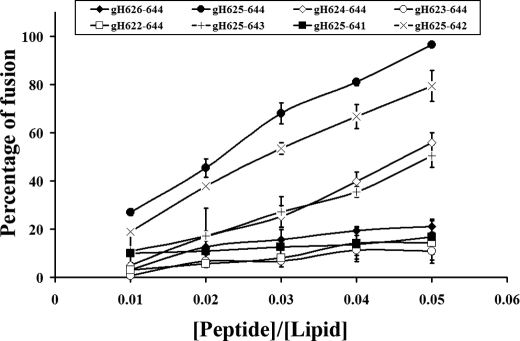
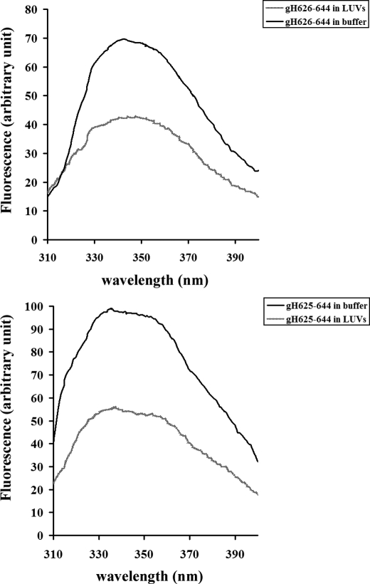
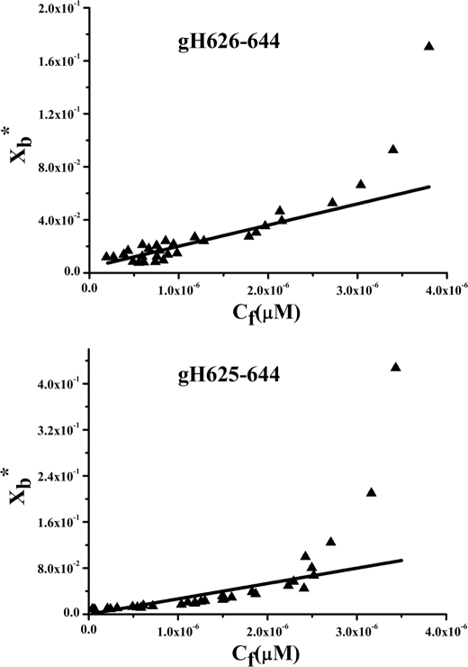
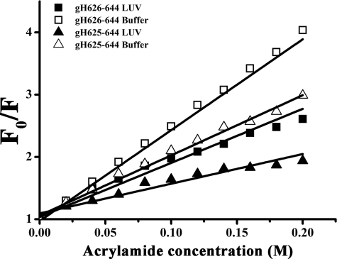
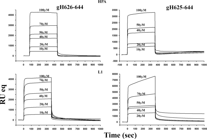
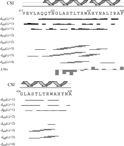
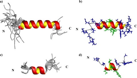
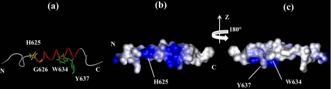
Herpes simplex virus type 1 (HSV-1)-induced membrane fusion remains one of the most elusive mechanisms to be deciphered in viral entry. The structure resolution of glycoprotein gB has revealed the presence of fusogenic domains in this protein and pointed out the key role of gB in the entry mechanism of HSV-1. A second putative fusogenic glycoprotein is represented by the heterodimer comprising the membrane-anchored glycoprotein H (gH) and the small secreted glycoprotein L, which remains on the viral envelope in virtue of its non-covalent interaction with gH. Different domains scattered on the ectodomain of HSV-1 gH have been demonstrated to display membranotropic characteristics. The segment from amino acid 626 to 644 represents the most fusogenic region identified by studies with synthetic peptides and model membranes. Herein we have identified the minimal fusogenic sequence present on gH. An enlongation at the N terminus of a single histidine (His) has proved to profoundly increase the fusogenic activity of the original sequence. Nuclear magnetic resonance (NMR) studies have shown that the addition of the N-terminal His contributes to the formation and stabilization of an alpha-helical domain with high fusion propensity.
Figures
Similar articles
-
Role of membranotropic sequences from herpes simplex virus type I glycoproteins B and H in the fusion process.Biochim Biophys Acta. 2010 Mar;1798(3):579-91. doi: 10.1016/j.bbamem.2010.01.006. Epub 2010 Jan 18. Biochim Biophys Acta. 2010. PMID: 20085747
-
Structure and orientation of the gH625-644 membrane interacting region of herpes simplex virus type 1 in a membrane mimetic system.Biochemistry. 2012 Apr 10;51(14):3121-8. doi: 10.1021/bi201589m. Epub 2012 Mar 23. Biochemistry. 2012. PMID: 22397737
-
Analysis of a membrane interacting region of herpes simplex virus type 1 glycoprotein H.J Biol Chem. 2008 Oct 31;283(44):29993-30009. doi: 10.1074/jbc.M803092200. Epub 2008 Aug 4. J Biol Chem. 2008. PMID: 18678872 Free PMC article.
-
Understanding HSV-1 entry glycoproteins.Rev Med Virol. 2007 May-Jun;17(3):205-15. doi: 10.1002/rmv.531. Rev Med Virol. 2007. PMID: 17295428 Free PMC article. Review.
-
Molecular gymnastics at the herpesvirus surface.EMBO Rep. 2006 Oct;7(10):1000-5. doi: 10.1038/sj.embor.7400807. EMBO Rep. 2006. PMID: 17016458 Free PMC article. Review.
Cited by
-
Eradication of Candida albicans persister cell biofilm by the membranotropic peptide gH625.Sci Rep. 2020 Apr 1;10(1):5780. doi: 10.1038/s41598-020-62746-w. Sci Rep. 2020. PMID: 32238858 Free PMC article.
-
Liposome armed with herpes virus-derived gH625 peptide to overcome doxorubicin resistance in lung adenocarcinoma cell lines.Oncotarget. 2016 Jan 26;7(4):4077-92. doi: 10.18632/oncotarget.6013. Oncotarget. 2016. PMID: 26554306 Free PMC article.
-
Peptides and Dendrimers: How to Combat Viral and Bacterial Infections.Pharmaceutics. 2021 Jan 14;13(1):101. doi: 10.3390/pharmaceutics13010101. Pharmaceutics. 2021. PMID: 33466852 Free PMC article. Review.
-
Antimicrobial Dendrimeric Peptides: Structure, Activity and New Therapeutic Applications.Int J Mol Sci. 2017 Mar 3;18(3):542. doi: 10.3390/ijms18030542. Int J Mol Sci. 2017. PMID: 28273806 Free PMC article. Review.
-
Peptide gH625 enters into neuron and astrocyte cell lines and crosses the blood-brain barrier in rats.Int J Nanomedicine. 2015 Mar 10;10:1885-98. doi: 10.2147/IJN.S77734. eCollection 2015. Int J Nanomedicine. 2015. PMID: 25792823 Free PMC article.
References
-
- Brasseur R., Pillot T., Lins L., Vandekerckhove J., Rosseneu M. (1997) Trends Biochem. Sci. 22, 167–171 - PubMed
MeSH terms
Substances
LinkOut - more resources
Full Text Sources
Other Literature Sources
