. 2011 Feb;44(2):155-65.
doi: 10.1165/rcmb.2009-0454OC. Epub 2010 Mar 26.

Inhibition by cigarette smoke of nuclear factor-κB-dependent response to bacteria in the airway

Affiliations

Inhibition by cigarette smoke of nuclear factor-κB-dependent response to bacteria in the airway

Lori J Manzel et al. Am J Respir Cell Mol Biol. 2011 Feb.

Abstract

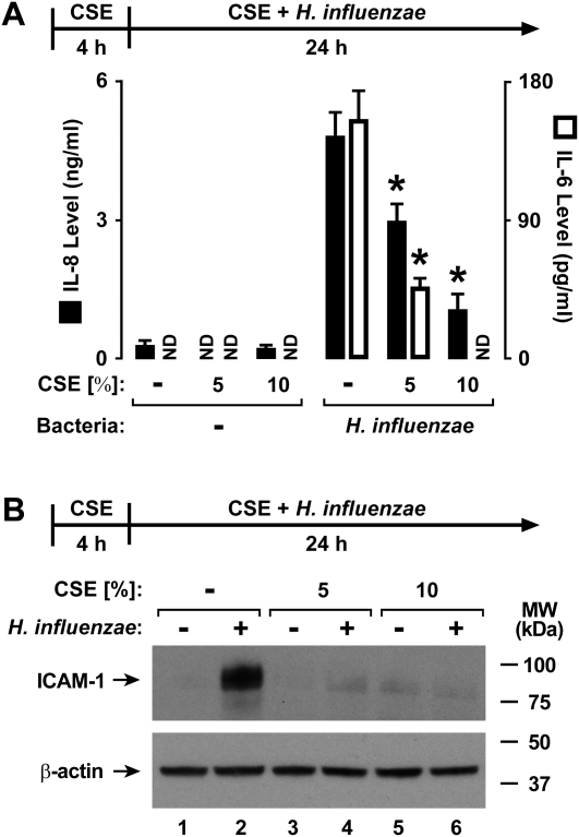
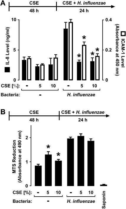
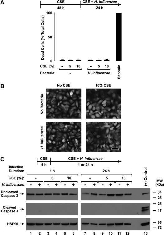
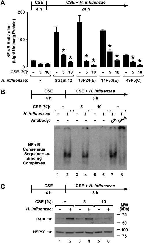
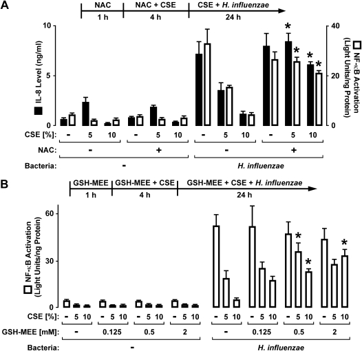
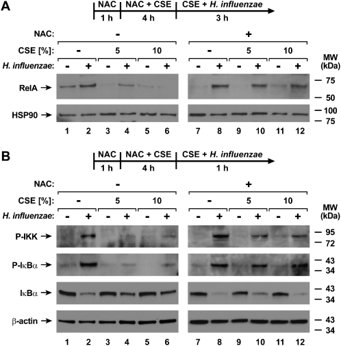
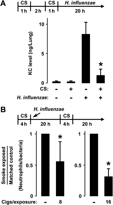
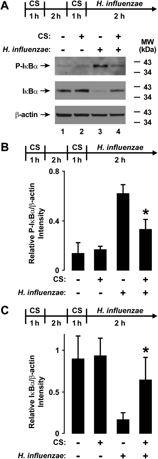
Although individuals exposed to cigarette smoke are more susceptible to respiratory infection, the effects of cigarette smoke on pulmonary defense are incompletely understood. Based on the observation that interactions between bacteria and host cells result in the expression of critical defense genes regulated by NF-κB, we hypothesized that cigarette smoke alters NF-κB function. In this study, primary human tracheobronchial epithelial cells were treated with cigarette smoke extract (CSE) and exposed to Haemophilus influenzae, and the effects of CSE on bacteria-induced signaling and gene expression were assessed. CSE inhibited high concentrations of induced NF-κB activation and the consequent expression of defense genes that occurred in airway epithelial cells in response to H. influenzae. This decreased activation of NF-κB was not attributable to cell loss or cytotoxicity. Glutathione augmentation of epithelial cells decreased the effects of CSE on NF-κB-dependent responses, as well as the effects on the inhibitor of κB and the inhibitor of κB kinase, which are upstream NF-κB regulators, suggesting the involvement of reactive oxygen species. The relevance of these findings for lung infection was confirmed using a mouse model of H. influenzae airway infection, in which decreased NF-κB pathway activation, keratinocyte chemoattractant (KC) chemokine expression, and neutrophil recruitment occurred in animals exposed to cigarette smoke. The results indicate that although cigarette smoke can cause inflammation in the lung, exposure to smoke inhibits the robust pulmonary defense response to H. influenzae, thereby providing one explanation for the increased susceptibility to respiratory bacterial infection in individuals exposed to cigarette smoke.


Figures

Figure 1.
Figure 1.
Cigarette smoke extract (CSE) decreases Haemophilus influenzae–induced defense protein expression. (A) IL-8 and IL-6 secretion levels in culture medium were determined using enzyme-linked immunoassays with human tracheobronchial epithelial (hTBE) cells that were first treated with medium containing CSE at the indicated concentrations for 4 hours, followed by incubation for 24 hours in medium containing the same CSE concentration with or without H. influenzae strain 12. Values are expressed as mean ± SD (n = 3). *Significant difference in concentrations from cells treated with and without CSE. ND, none detected. (B) Intercellular adhesion molecule–1 (ICAM-1) and β-actin protein concentrations were assessed using immunoblot analyses of extracts from hTBE cells that were first treated with medium containing CSE at the indicated concentrations for 4 hours, followed by incubation for 24 hours in medium containing the same CSE concentrations with or without H. influenzae strain 12. MW, molecular weight.
Figure 2.
Figure 2.
CSE decreases H. influenzae–induced defense protein and mRNA expression. (A) ICAM-1 cell surface protein concentrations were determined using an enzyme-linked immunoassay with hTBE cell monolayers that were first treated with medium containing CSE at the indicated concentrations for 4 hours. Cells were then incubated for 24 hours in medium containing the same CSE concentration with or without H. influenzae strain 12 or strains from patients with chronic obstructive pulmonary disease (COPD) isolated from sputum during an exacerbation (E) or that colonized during symptom baseline (C). (B) IL-8 and ICAM-1 mRNA concentrations were determined using real-time RT-PCR analyses of total RNA from hTBE cells that were first treated with medium containing CSE at the indicated concentrations for 4 hours, followed by incubation for 4 hours in medium containing the same CSE concentrations with or without H. influenzae strain 12. Values are expressed as mean relative mRNA concentrations compared with control human hypoxanthine phosphoribosyltransferase (HPRT) mRNA. Values are expressed as mean ± SD (n = 3).*Significant difference in concentrations from cells treated with or without CSE.
Figure 3.
Figure 3.
CSE selectively alters the effects of H. influenzae on airway epithelial cells. (A) IL-8 secretion into culture medium and ICAM-1 cell-surface protein concentrations were determined using enzyme-linked immunoassays with hTBE cells that were first treated with medium containing CSE at the indicated concentrations for 48 hours, followed by incubation for 24 hours in medium containing the same CSE concentrations with or without H. influenzae strain 12. (B) Mitochondrial activity was determined using a 3-(,5-dimethylthiazol-2-yl)-5-(3-carboxymethoxyphenyl)-2-(4-sulfophenyl)-2H-tetrazolium (MTS)–based assay with hTBE cells that were first treated with medium containing CSE at the indicated concentrations for 48 hours, followed by incubation for 24 hours in medium containing the same CSE concentrations, with or without H. influenzae strain 12. Control samples were treated with 1% saponin to induce 100% cell death. Values are expressed as mean ± SD (n = 3). *Significant difference in concentrations from cells treated without compared with CSE.
Figure 4.
Figure 4.
CSE causes minimal airway epithelial cell cytotoxicity. (A and B) Dead and live cell numbers were quantified using hTBE cells that were first treated with medium containing CSE at the indicated concentrations for 48 hours, followed by incubation for 24 hours in medium containing the same CSE concentrations, with or without H. influenzae strain 12. Plasma membrane permeability to ethidium homodimers was detected in dead cells, and intracellular esterase activity was identified in live cells. (A) Control samples were treated with 1% saponin to induce 100% cell death. Values were calculated as mean percent dead cells/total cells ± SD for four random low-power fields (∼ 500–750 cells/field) from duplicate samples. No significant difference between conditions was evident. (B) Photomicrographs show cell morphology after cell treatments and esterase activity staining in this assay. Bar, 50 μm. (C) Uncleaved and cleaved caspase-3 and heat shock protein (HSP)–90 protein concentrations were assessed using immunoblot analyses of extracts from hTBE cells that were first treated with medium containing CSE at the indicated concentrations for 4 hours, followed by incubation for 1 hour or 24 hours in medium containing the same CSE concentrations, with or without H. influenzae strain 12. As a positive control for caspase cleavage, Jurkat cell extract was treated with cytochrome c (Cell Signaling Technology).
Figure 5.
Figure 5.
CSE inhibits H. influenzae–induced NF-κB activation. (A) NF-κB–dependent reporter gene activation was assessed using luciferase activity assays of extracts from hTBE cells that were initially infected with an adenoviral vector expressing a luciferase gene driven by four tandem NF-κB sites. Cells were then treated with medium containing CSE at the indicated concentrations for 4 hours, followed by incubation for 24 hours in medium containing the same CSE concentrations, with or without H. influenzae strain 12 or strains from patients with COPD isolated from sputum during an exacerbation (E) or that colonized during symptom baseline (C). Values are expressed as mean ± SD (n = 3–6). *Significant difference in concentrations from cells treated with or without CSE. (B) Transcription factor binding to an NF-κB consensus sequence was assessed using electrophoretic mobility-shift assay (EMSA) with nuclear extracts from hTBE cells that were first treated with medium containing CSE at the indicated concentrations for 4 hours, followed by incubation for 3 hours in medium containing the same CSE concentrations, with or without H. influenzae strain 12. Supershift analysis was performed by the addition of control antibodies or antibodies against NF-κB RelA. (C) NF-κB RelA and HSP-90 nuclear protein concentrations in hTBE cells were assessed using immunoblot analyses of nuclear extracts from B.
Figure 6.
Figure 6.
Glutathione augmentation inhibits effects of CSE on airway epithelial cells. (A) IL-8 secretion concentration in hTBE cell culture medium was determined using an enzyme-linked immunoassay. NF-κB–dependent reporter gene activation was assessed using the reporter gene adenoviral vector and luciferase activity assays of hTBE cell extracts. Cells were treated with medium containing CSE at the indicated concentrations for 4 hours, followed by incubation for 24 hours in medium containing the same CSE concentrations, with or without H. influenzae strain 12. Some samples were also incubated with 20 mM N-acetylcysteine (NAC) from 1 hour before the addition of CSE until incubation with bacteria. (B) NF-κB–dependent reporter gene activation was assessed using the reporter gene adenoviral vector and luciferase activity assays of extracts from hTBE cells that were treated with medium containing CSE at the indicated concentrations for 4 hours, followed by incubation for 24 hours in medium containing the same CSE concentrations, with or without H. influenzae strain 12. Some samples were also incubated with the indicated concentrations of glutathione monoethyl ester (GSH-MEE) from 1 hour before the addition of CSE until the end of the experiment. Values are expressed as mean ± SD (n = 3). *Significant difference in comparable samples treated with or without NAC or GSH-MEE.
Figure 7.
Figure 7.
Glutathione augmentation inhibits effects of CSE on H. influenzae–induced cell signaling. (A) NF-κB RelA and HSP-90 nuclear protein concentrations were assessed using immunoblot analyses of nuclear extracts from hTBE cells that were treated with medium containing CSE at the indicated concentrations for 4 hours, followed by incubation for 3 hours in medium containing the same concentrations of CSE, with or without H. influenzae strain 12. (B) Phosphorylated (P) inhibitor of κB kinase (IKK) and of κB-inhibitor (IκB)–α, total IκBα, and β-actin protein concentrations were assessed using immunoblot analyses of extracts from hTBE cells that were treated with medium containing CSE at the indicated concentrations for 4 hours, followed by incubation for 1 hour in medium containing the same concentrations of CSE, with or without H. influenzae strain 12. Some samples were also incubated with 20 mM NAC from 1 hour before the addition of CSE until incubation with bacteria.
Figure 8.
Figure 8.
Cigarette smoke modulates in vivo airway antibacterial defense. (A) keratinocyte chemoattractant (KC) concentrations were determined using an enzyme-linked immunoassay with lung extracts from C57BL/6J mice that were sham-exposed or exposed to cigarette smoke (CS) for 2 out of 4 hours. Whole-lung samples were isolated 20 hours after endotracheal injection with agar particles combined with H. influenzae strain 12. Values are expressed as mean ± SD (n = 3–5 in each group). *Significant difference between bacterial infection with or without exposure to CS. (B) The ratio of bronchoalveolar lavage neutrophil number to lung bacterial load was determined in C57BL/6J mice that were sham-exposed or exposed to eight or 16 cigarettes over 4 hours, followed by endotracheal injection with agar particles combined with H. influenzae strain 12. After 20 hours, animals underwent a second exposure to CS, and lungs were isolated after 44 hours of infection. Smoke-exposed and control animals were matched by approximately equivalent bacterial loads, and animals were exposed to either eight or 16 cigarettes in independent experiments. Values are expressed as mean smoke-exposed/control animal ratio ± SD (n = 7 in each group). *Significant difference between animals with or without exposure to CS.
Figure 9.
Figure 9.
CS inhibits H. influenzae–induced NF-κB pathway signaling in vivo. (A) Phosphorylated and total IκBα and β-actin protein concentrations were assessed using immunoblot analyses of lung extracts from C57BL/6J mice that were sham-exposed or exposed to CS for 2 out of 4 hours. Whole-lung samples were isolated 2 hours after an endotracheal injection with agar particles combined with H. influenzae strain 12. (B) Whole-lung phosphorylated IκBα protein concentrations in A were quantified using results from band densitometry of immunoblot analyses, with inclusion of samples from additional animals. (C) Whole-lung total IκBα protein concentrations in A were quantified using results from band densitometry of immunoblot analyses, with inclusion of samples from additional animals. In B and C, values are expressed as mean ± SD (n = 3–5 in each group). *Significant difference between bacterial infection with or without exposure to CS.

Similar articles

Cited by

References

    1. Brook I, Gober AE. Recovery of potential pathogens and interfering bacteria in the nasopharynx of smokers and nonsmokers. Chest 2005;127:2072–2075. - PubMed
    1. Nuorti JP, Butler JC, Farley MM, Harrison LH, McGeer A, Kolczak MS, Breiman RF. Cigarette smoking and invasive pneumococcal disease: Active Bacterial Core Surveillance Team. N Engl J Med 2000;342:681–689. - PubMed
    1. US Department of Health and Human Services. The health consequences of involuntary exposure to tobacco smoke: a report of the surgeon general. Atlanta: Department of Health and Human Services, Centers for Disease Control and Prevention, Coordinating Center for Health Promotion, National Center for Chronic Disease Prevention and Health Promotion, Office on Smoking and Health; 2006.
    1. Sopori M. Effects of cigarette smoke on the immune system. Nat Rev Immunol 2002;2:372–377. - PubMed
    1. Stanley PJ, Wilson R, Greenstone MA, MacWilliam L, Cole PJ. Effect of cigarette smoking on nasal mucociliary clearance and ciliary beat frequency. Thorax 1986;41:519–523. - PMC - PubMed

Publication types

MeSH terms