. 2010 May;176(5):2283-91.
doi: 10.2353/ajpath.2010.091069. Epub 2010 Mar 26.

Human intestinal ischemia-reperfusion-induced inflammation characterized: experiences from a new translational model

Affiliations

Human intestinal ischemia-reperfusion-induced inflammation characterized: experiences from a new translational model

Joep Grootjans et al. Am J Pathol. 2010 May.

Abstract

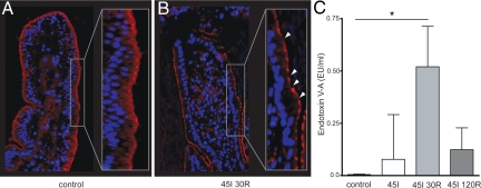
Human intestinal ischemia-reperfusion (IR) is a frequent phenomenon carrying high morbidity and mortality. Although intestinal IR-induced inflammation has been studied extensively in animal models, human intestinal IR induced inflammatory responses remain to be characterized. Using a newly developed human intestinal IR model, we show that human small intestinal ischemia results in massive leakage of intracellular components from ischemically damaged cells, as indicated by increased arteriovenous concentration differences of intestinal fatty acid binding protein and soluble cytokeratin 18. IR-induced intestinal barrier integrity loss resulted in free exposure of the gut basal membrane (collagen IV staining) to intraluminal contents, which was accompanied by increased arteriovenous concentration differences of endotoxin. Western blot for complement activation product C3c and immunohistochemistry for activated C3 revealed complement activation after IR. In addition, intestinal IR resulted in enhanced tissue mRNA expression of IL-6, IL-8, and TNF-alpha, which was accompanied by IL-6 and IL-8 release into the circulation. Expression of intercellular adhesion molecule-1 was markedly increased during reperfusion, facilitating influx of neutrophils into IR-damaged villus tips. In conclusion, this study for the first time shows the sequelae of human intestinal IR-induced inflammation, which is characterized by complement activation, production and release of cytokines into the circulation, endothelial activation, and neutrophil influx into IR-damaged tissue.


Figures

Figure 1
Figure 1
Intestinal IR-induced epithelial lining breakdown resulting in exposure of the basal membrane to intraluminal content. Hematoxylin and eosin staining (×200) shows an intact villus structure in control tissue (A), whereas 45I led to disruption of the epithelial lining (arrowheads) and appearance of subepithelial spaces (arrows) (B). After 45I-30R, damaged enterocytes were shed into the lumen (C). The epithelial lining remained compromised after 45I-120R (D). Collagen IV staining (red) revealed that enterocytes were attached to the basal membrane in control tissue (E). Destruction of the epithelial lining after 45I and 45I-30R resulted in exposure of the basal membrane to intraluminal contents (F and G respectively, arrows), which was not fully reversed after 45I-120R (H).
Figure 2
Figure 2
Intestinal IR results in apoptosis and massive leakage of intracellular components from IR-damaged enterocytes. A: I-FABP arteriovenous concentration differences increased on reperfusion (***P < 0.001), reflecting enterocyte membrane integrity loss. This was accompanied by leakage of M65 from dying cells (**P < 0.01). Both I-FABP and M65 levels decreased over the course of reperfusion. Arteriovenous concentration differences of M30 did not change over time. B: Immunohistochemistry for M30 showed that apoptosis was sporadically present in villus tips of healthy jejunum and jejunum exposed to ischemia alone (left and right upper, respectively). M30 positivity was mainly observed in detaching enterocytes at 30 minutes of reperfusion (left lower) and in the luminal debris of IR-damaged cells at 120 minutes of reperfusion (right lower).
Figure 3
Figure 3
Intestinal IR-induced tight junction loss results in translocation of endotoxin to the circulation. Immunofluorescence for ZO-1 shows continuous staining in control jejunal tissue (A), whereas an interrupted staining pattern was observed over the villus lining after 45 minutes of ischemia with 30 minutes reperfusion (arrowheads), indicating tight junction loss (B). C: Significant translocation of endotoxin into the circulation was observed at 45 minutes of ischemia with 30 minutes of reperfusion (*P < 0.05).
Figure 4
Figure 4
Activation of the complement system in response to intestinal IR. A: Western blot analysis revealed that C3c, a product of C3 activation, was abundantly present in the luminal debris of shedded cells, whereas low C3c levels were observed in tissue homogenates of normal and IR-exposed jejunum (AU = arbitrary units, ***P < 0.001) B: In contrast, native C3 (113-kD bands) was present in both normal jejunum and jejunum exposed to IR, whereas it was hardly detected in luminal debris of IR-damaged, shedded cells. (AU indicates arbitrary units, **P < 0.01) C: Immunohistochemistry revealed the presence of activated C3 in luminal debris after 45 minutes of ischemia with 120 minutes of reperfusion.
Figure 5
Figure 5
Endothelial activation and influx of neutrophils into IR-damaged intestinal tissue. A: ICAM-1 staining reveals endothelial activation after 45 minutes of ischemia with 30 and 120 minutes of reperfusion, which was accompanied by influx of neutrophils into IR-damaged villus tips, as shown by MPO staining. B: The number of MPO-positive cells was significantly increased in IR-damaged villi after 30 and 120 minutes of reperfusion (***P < 0.001 and *P < 0.05, respectively).
Figure 6
Figure 6
Intestinal IR results in increased IL-6, IL-8, and TNF-α mRNA tissue expression, which is accompanied by release of IL-6 and IL-8 into the circulation. IL-6 (A) and IL-8 (B) mRNA expression and protein release into the circulation increased significantly during reperfusion of the ischemically damaged jejunal segment. In addition, intestinal IR resulted in increased mRNA expression of TNF-α (C). *P < 0.05, **P < 0.01, ***P < 0.001.

Similar articles

Cited by

References

    1. American Gastroenterological Association Medical Position Statement: guidelines on intestinal ischemia. Gastroenterology. 2000;118:951–953. - PubMed
    1. Chen Y, Lui VC, Rooijen NV, Tam PK. Depletion of intestinal resident macrophages prevents ischaemia reperfusion injury in gut. Gut. 2004;53:1772–1780. - PMC - PubMed
    1. Blikslager AT, Moeser AJ, Gookin JL, Jones SL, Odle J. Restoration of barrier function in injured intestinal mucosa. Physiol Rev. 2007;87:545–564. - PubMed
    1. Kubes P, Hunter J, Granger DN. Ischemia/reperfusion-induced feline intestinal dysfunction: importance of granulocyte recruitment. Gastroenterology. 1992;103:807–812. - PubMed
    1. Homer-Vanniasinkam S, Crinnion JN, Gough MJ. Post-ischaemic organ dysfunction: a review. Eur J Vasc Endovasc Surg. 1997;14:195–203. - PubMed

Publication types