. 2010 May;176(5):2490-9.
doi: 10.2353/ajpath.2010.090777. Epub 2010 Mar 26.

Contribution of MyD88 to the tumor exosome-mediated induction of myeloid derived suppressor cells

Affiliations

Contribution of MyD88 to the tumor exosome-mediated induction of myeloid derived suppressor cells

Yuelong Liu et al. Am J Pathol. 2010 May.

Abstract

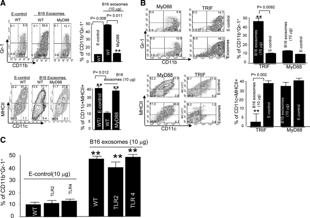
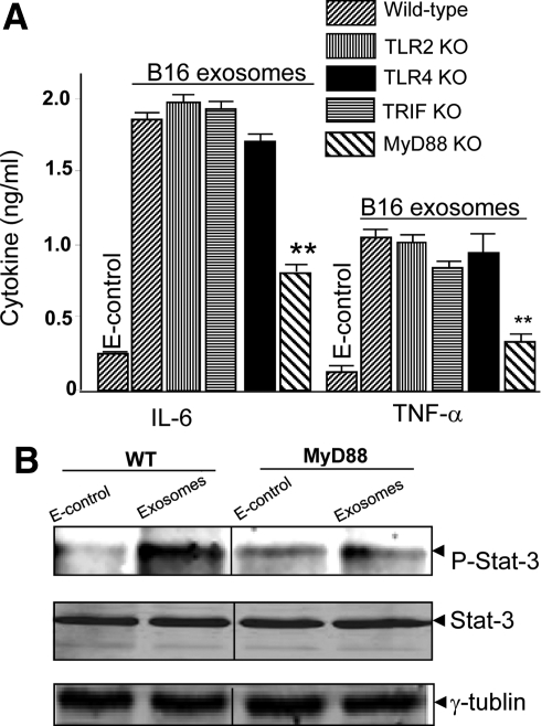
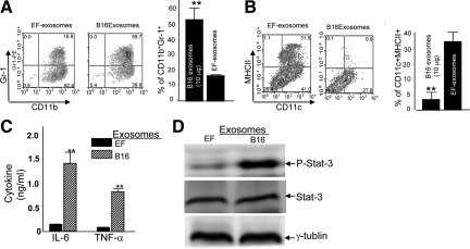
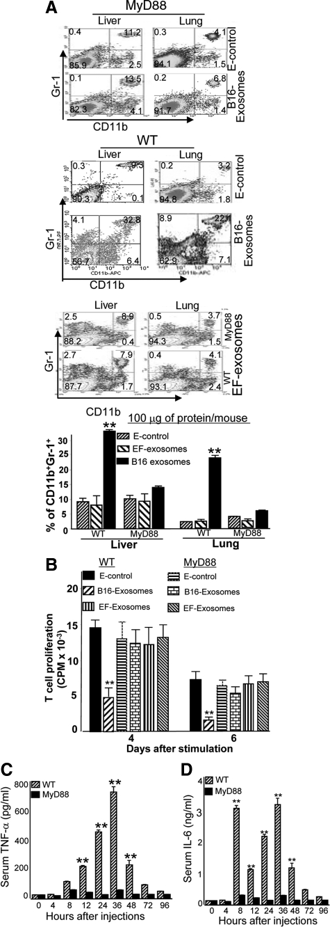
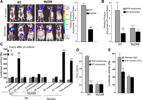
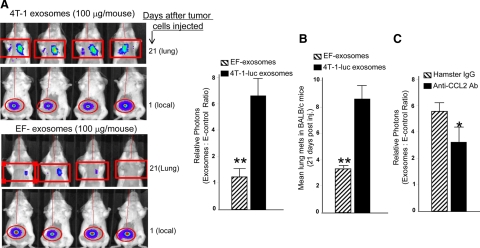
In this study we observed that mice pretreated with tumor exosomes had a significant acceleration of tumor metastasis in the lung. Tumor metastasis correlated significantly with an increase in recruitment of more Myeloid-derived suppressor cells (MDSCs) in the lung of C57BL/6j (B6) mice pretreated with tumor exosomes. These effects were blunted when MyD88 knockout (KO) mice were pretreated with tumor exosomes. MDSCs induced by tumor exosomes and isolated from wild-type B6 mice also more potently inhibited T cell activation and induction of interleukin-6 and tumor necrosis factor-alpha than MDSCs isolated from the lung of MyD88 KO mice. In vitro, addition of tumor exosomes to bone marrow-derived CD11b(+)Gr-1(+) cells isolated from wild-type B6 mice resulted in more cytokine production, including tumor necrosis factor-alpha, interleukin-6, and the chemokine CCL2, than CD11b(+)Gr-1(+) cells isolated from MyD88 KO mice. Moreover, lower levels of CCL2 were observed in the lungs in MyD88 KO mice pretreated with tumor exosomes than that in wild-type mice. Together these data demonstrate a pivotal role for MyD88 in tumor exosome-mediated expansion of MDSCs and tumor metastasis.


Figures

Figure 1
Figure 1
MyD88 dependent and tumor exosome-mediated blocking of the differentiation of GM-CSF-stimulated BM precursor cells. Erythrocyte-depleted BM cells of wild-type B6 mice or B6 mice with specific gene knockouts listed in A and B, were cultured in RPMI 1640 supplemented with 10% FBS and 20 ng/ml recombinant mouse GM-CSF. B16 exosomes or E-control (10 μg/ml) were added at day 0 after the addition of the GM-CSF to the cell cultures (AC). After seven days in culture, the cells were analyzed by FACS for the expression of CD11b, Gr-1, CD11c, and MHCII. One representative of five independent experiments is shown (AC). All data are given as the average values ± SEM obtained for five samples in three independent experiments (AC). **P < 0.01.
Figure 2
Figure 2
MyD88 dependent induction of proinflammatory cytokines and stat3 activation. Supernatants of two-day cultures treated as described in Figure 1, A–C, were collected, and the amount of interleukin-6 (A; left) and TNF-α (A; right) was measured by using a standard ELISA. **P < 0.01. To determine the effect of MyD88 on B16 exosome-mediated induction of phosphorylated Stat3, CD11b+ cells were treated with B16 exosomes or E-control (10 μg/ml), and phosphorylated Stat3 was detected in 48-hour cultured cells by Western blot analysis (B). Total cell lysates (100 μg/lane) were also used for determination of total Stat3 (B; middle) or γ-tubulin (B; bottom) by Western blot analysis.
Figure 3
Figure 3
Tumor exosome specific-mediated blocking of the differentiation of GM-CSF-stimulated BM precursor cells. B16 exosomes or EF-exosomes (10 μg/ml) were added at day 0 after the addition of the GM-CSF to the cell cultures (AC). Exosomal effects on the production of CD11b+Gr-1+ cells (A), dendritic cells (B), interleukin-6 and TNF-α (C), and activation of Stat3 (D) were evaluated by using identical procedures as described in Figures 1 and 2. Data represent mean ± SEM of three samples in three independent experiments. **P < 0.01.
Figure 4
Figure 4
Intravenous injection of tumor exosomes leads to the induction of MDSCs via a MyD88-mediated pathway. A total of 100 μg of B16 exosomes, EF-exosomes, or E-control in 100 μl of PBS was injected into MyD88 knockout and B6 wild-type mice via the tail vein twice a week for three weeks. One day after the last injection, the mice were sacrificed, and the percentages of CD11b+Gr-1+ cells in lung and liver tissues were determined by FACS analysis (A). CD11b+Gr-1+ cells (5 × 10⁵) were isolated from the lung by FACS sorting as described in the Materials and Methods, and the isolated cells were co-cultured with splenic T cells at a ratio of 5:1 (B). Cell proliferation was measured by [3H]thymidine incorporation after four and six days in culture. Data are presented as the mean ± SEM of triplicate wells representing three independent experiments. B6 wild-type or MyD88 mice were intravenously injected with B16 exosomes (100 μg/mouse in 100 μl of PBS). Sera were collected up to 96 hours after injection (C and D) and analyzed for TNF-α (C) and interleukin-6 (D) by using an ELISA. Data represent mean ± SEM of five mouse sera in two independent experiments. **P < 0.01.
Figure 5
Figure 5
MyD88 adaptive molecule plays a role in tumor exosome-mediated promotion of B16 tumor cell growth in the lung and in tumor chemotaxis. Seven-week-old wild-type B6 or MyD88 knockout mice were injected intravenously with B16 exosomes (100 μg/mouse in 100 μl of PBS) or E-control twice a week for three weeks. One day after the last injection, the mice were injected intravenously with B16–luc cells (1 × 10⁵). Twenty-one days after B16-luc injection, the mice were imaged at 0 and 4 hours (A) after injection of D-luciferin, and the total photon count per minute (photons per minute) was calculated (nine animals/group, three representative mice are shown) by using Living Image software (A; left). The growth potential of injected B16–luc cells was determined by dividing photon emissions at 4 hours by the photon emissions at 0 hours (A; right) for mice treated with exosomes or the E-control. To determine the specificity of B16 exosome-mediated promotion of B16-luc growth in the lung, mice were treated with EF-exosomes or B16 exosomes by using an identical protocol as described in A. In vivo luciferase activity was measured as described in A. All results in B are based on two independent experiments with data pooled for mice in each experiment. Results in both A and B are presented as the mean ± SEM; **P < 0.01. Seven-day cultured BM precursor cells isolated from B6 wild-type mice or BALB/c mice and treated as described in Figure 2A were co-cultured with B16-luc or 4T-1-luc in a 24-well transwell plate. Twenty-four hours later the cells in the bottom chamber were harvested, lysed, and luciferase activity was determined. Results are presented as percent of luciferase activity. Data presented are mean ± SEM of triplicates wells for three independent experiments (C). Supernatants of seven-day cultures treated as described in Figure 1, A–C, were collected, and the amount of CCL2 (D) was measured by using a standard ELISA. Data represent mean ± SEM of three samples in three independent experiments. **P < 0.01. Seven-day cultured BM precursor cells treated as described in Figure 2A were co-cultured with B16-luc in the presence of either anti-mouse CCL2 (1.0 μg/ml) or a control hamster IgG in a 24-well transwell plate. Twenty-four hours later the cells in the bottom chamber were harvested, lysed, and luciferase activity was determined. Results are presented as percent of luciferase activity. Data presented are mean ± SEM of triplicates wells for three independent experiments (E).
Figure 6
Figure 6
Tumor exosome-mediated promotion of 4T-1 tumor metastasis in the lung. A: A total of 100 μg of 4T-1-luc exosomes or EF-exosomes in 100 μl of PBS was injected into BALB/c female mice via the tail vein twice a week for three weeks. One day after the last injection, 1 × 10⁵ 4T-1-luc cells were injected in the right flank of each mouse. The next day, to verify that approximately equal numbers of tumor cells were injected, mice were imaged by using a luciferase imaging assay as described in Figure 5A. A representative image from each group of mice is shown (A; top panels of each group). Ten days after 4T-1-luc injection, tumors were removed. Twenty-one days after tumor cells were injected, in vivo luciferase activity in the lung was determined by using an identical protocol as described in Figure 5A. Representative images of mice are shown (A; bottom panels of each group). The results are based on two independent experiments with data pooled for eight mice in each experiment. Results are presented as the mean ± SEM. **P < 0.01 (A; bar graph). Lung metastases on stained slides were counted manually by using a microscope, and the mean number per section was plotted (B). Statistical significance was analyzed by the nonparametric Mann Whitney t-test. Mice were treated with exosomes as described in A, and then identical protocols as described in Figure 5A were used for 4T-1-luc tumor cell injection and tumor removal. One day after tumor cells were injected, mice were intravenously injected at dose 10 mg/kg of body weight of mouse with hamster anti-CCL2 antibody or a control hamster IgG antibody every four days until the end of the mouse experiment. Mice were imaged at day 21 after the tumor cells were injected. The results are based on two independent experiments with data pooled for eight mice in each experiment. Results are presented as the mean ± SEM (C). *P < 0.05.

References

    1. Coussens LM, Werb Z. Inflammation and cancer. Nature. 2002;420:860–867. - PMC - PubMed
    1. Lee JJ, Lotze MT. Molecular basis of metastasis. N Engl J Med. 2009;360:1679. author reply 1679–1680. - PubMed
    1. Melnikova VO, Bar-Eli M. Inflammation and melanoma metastasis. Pigment Cell Melanoma Res. 2009;22:257–267. - PubMed
    1. Mon NN, Kokuryo T, Hamaguchi M. Inflammation and tumor progression: a lesson from TNF-alpha-dependent FAK signaling in cholangiocarcinoma. Methods Mol Biol. 2009;512:279–293. - PubMed
    1. Yu S, Liu C, Su K, Wang J, Liu Y, Zhang L, Li C, Cong Y, Kimberly R, Grizzle WE, Falkson C, Zhang HG. Tumor exosomes inhibit differentiation of bone marrow dendritic cells. J Immunol. 2007;178:6867–6875. - PubMed

Publication types