

ArcS, the cognate sensor kinase in an atypical Arc system of Shewanella oneidensis MR-1
- PMID: 20348304
- PMCID: PMC2869118
- DOI: 10.1128/AEM.00512-10
ArcS, the cognate sensor kinase in an atypical Arc system of Shewanella oneidensis MR-1
Abstract
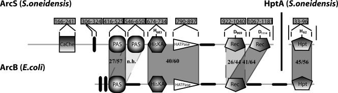
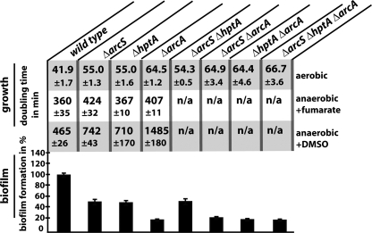
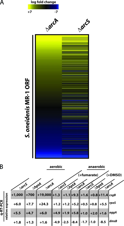
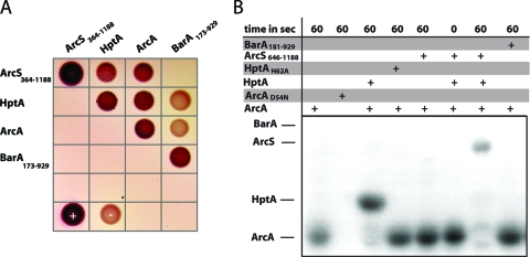
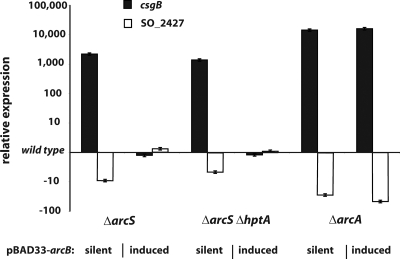
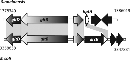
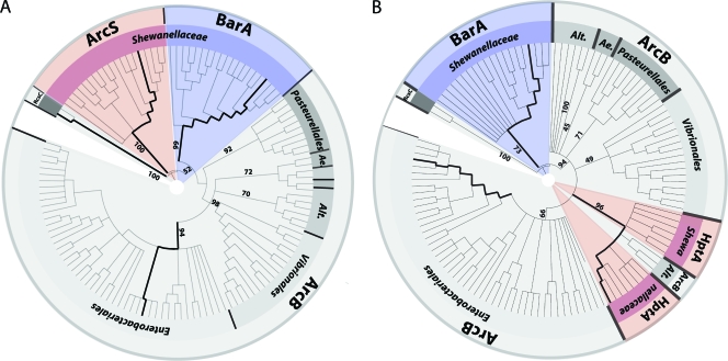
The availability of oxygen is a major environmental factor for many microbes, in particular for bacteria such as Shewanella species, which thrive in redox-stratified environments. One of the best-studied systems involved in mediating the response to changes in environmental oxygen levels is the Arc two-component system of Escherichia coli, consisting of the sensor kinase ArcB and the cognate response regulator ArcA. An ArcA ortholog was previously identified in Shewanella, and as in Escherichia coli, Shewanella ArcA is involved in regulating the response to shifts in oxygen levels. Here, we identified the hybrid sensor kinase SO_0577, now designated ArcS, as the previously elusive cognate sensor kinase of the Arc system in Shewanella oneidensis MR-1. Phenotypic mutant characterization, transcriptomic analysis, protein-protein interaction, and phosphotransfer studies revealed that the Shewanella Arc system consists of the sensor kinase ArcS, the single phosphotransfer domain protein HptA, and the response regulator ArcA. Phylogenetic analyses suggest that HptA might be a relict of ArcB. Conversely, ArcS is substantially different with respect to overall sequence homologies and domain organizations. Thus, we speculate that ArcS might have adopted the role of ArcB after a loss of the original sensor kinase, perhaps as a consequence of regulatory adaptation to a redox-stratified environment.
Figures
Similar articles
-
Domain analysis of ArcS, the hybrid sensor kinase of the Shewanella oneidensis MR-1 Arc two-component system, reveals functional differentiation of its two receiver domains.J Bacteriol. 2013 Feb;195(3):482-92. doi: 10.1128/JB.01715-12. Epub 2012 Nov 16. J Bacteriol. 2013. PMID: 23161031 Free PMC article.
-
Anaerobic regulation by an atypical Arc system in Shewanella oneidensis.Mol Microbiol. 2005 Jun;56(5):1347-57. doi: 10.1111/j.1365-2958.2005.04628.x. Mol Microbiol. 2005. PMID: 15882425
-
Probing regulon of ArcA in Shewanella oneidensis MR-1 by integrated genomic analyses.BMC Genomics. 2008 Jan 25;9:42. doi: 10.1186/1471-2164-9-42. BMC Genomics. 2008. PMID: 18221523 Free PMC article.
-
Signaling by the arc two-component system provides a link between the redox state of the quinone pool and gene expression.Antioxid Redox Signal. 2006 May-Jun;8(5-6):781-95. doi: 10.1089/ars.2006.8.781. Antioxid Redox Signal. 2006. PMID: 16771670 Review.
-
Cellular and molecular physiology of Escherichia coli in the adaptation to aerobic environments.J Biochem. 1996 Dec;120(6):1055-63. doi: 10.1093/oxfordjournals.jbchem.a021519. J Biochem. 1996. PMID: 9010748 Review.
Cited by
-
Impaired cell envelope resulting from arcA mutation largely accounts for enhanced sensitivity to hydrogen peroxide in Shewanella oneidensis.Sci Rep. 2015 May 15;5:10228. doi: 10.1038/srep10228. Sci Rep. 2015. PMID: 25975178 Free PMC article.
-
The Stand-Alone PilZ-Domain Protein MotL Specifically Regulates the Activity of the Secondary Lateral Flagellar System in Shewanella putrefaciens.Front Microbiol. 2021 Jun 1;12:668892. doi: 10.3389/fmicb.2021.668892. eCollection 2021. Front Microbiol. 2021. PMID: 34140945 Free PMC article.
-
A Proline-Rich Element in the Type III Secretion Protein FlhB Contributes to Flagellar Biogenesis in the Beta- and Gamma-Proteobacteria.Front Microbiol. 2020 Dec 15;11:564161. doi: 10.3389/fmicb.2020.564161. eCollection 2020. Front Microbiol. 2020. PMID: 33384667 Free PMC article.
-
Production of siderophores increases resistance to fusaric acid in Pseudomonas protegens Pf-5.PLoS One. 2015 Jan 8;10(1):e0117040. doi: 10.1371/journal.pone.0117040. eCollection 2015. PLoS One. 2015. PMID: 25569682 Free PMC article.
-
Division of labor and collective functionality in Escherichia coli under acid stress.Commun Biol. 2022 Apr 7;5(1):327. doi: 10.1038/s42003-022-03281-4. Commun Biol. 2022. PMID: 35393532 Free PMC article.
References
-
- Aiba, H., S. Adhya, and B. de Crombrugghe. 1981. Evidence for two functional gal promoters in intact Escherichia coli cells. J. Biol. Chem. 256:11905-11910. - PubMed
-
- Aiyar, A., Y. Xiang, and J. Leis. 1996. Site-directed mutagenesis using overlap extension PCR. Methods Mol. Biol. 57:177-191. - PubMed
Publication types
MeSH terms
Substances
Associated data
- Actions
LinkOut - more resources
Full Text Sources
Other Literature Sources
Molecular Biology Databases
Research Materials
