. 2010 Apr 13;107(15):6771-6.
doi: 10.1073/pnas.1001813107. Epub 2010 Mar 26.

Structural resolution of a tandem hormone-binding element in the insulin receptor and its implications for design of peptide agonists

Affiliations

Structural resolution of a tandem hormone-binding element in the insulin receptor and its implications for design of peptide agonists

Brian J Smith et al. Proc Natl Acad Sci U S A. .

Abstract

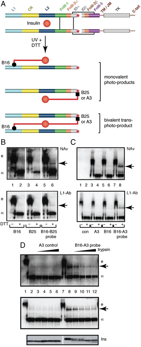
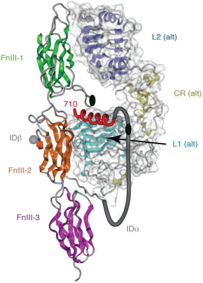
The C-terminal segment of the human insulin receptor alpha-chain (designated alphaCT) is critical to insulin binding as has been previously demonstrated by alanine scanning mutagenesis and photo-cross-linking. To date no information regarding the structure of this segment within the receptor has been available. We employ here the technique of thermal-factor sharpening to enhance the interpretability of the electron-density maps associated with the earlier crystal structure of the human insulin receptor ectodomain. The alphaCT segment is now resolved as being engaged with the central beta-sheet of the first leucine-rich repeat (L1) domain of the receptor. The segment is alpha-helical in conformation and extends 11 residues N-terminal of the classical alphaCT segment boundary originally defined by peptide mapping. This tandem structural element (alphaCT-L1) thus defines the intact primary insulin-binding surface of the apo-receptor. The structure, together with isothermal titration calorimetry data of mutant alphaCT peptides binding to an insulin minireceptor, leads to the conclusion that putative "insulin-mimetic" peptides in the literature act at least in part as mimics of the alphaCT segment as well as of insulin. Photo-cross-linking by novel bifunctional insulin derivatives demonstrates that the interaction of insulin with the alphaCT segment and the L1 domain occurs in trans, i.e., these components of the primary binding site are contributed by alternate alpha-chains within the insulin receptor homodimer. The tandem structural element defines a new target for the design of insulin agonists for the treatment of diabetes mellitus.


Conflict of interest statement

The authors declare no conflict of interest.

Figures

Fig. 1.
Fig. 1.
Structure of insulin and insulin receptor. (A) Alternate views of insulin monomer. A- and B-chains are shown in Light Gray and Dark Gray, respectively, with receptor-binding residues (1) in the A- and B-chains highlighted in Blue and Red, respectively. (B) Domain organization within the insulin receptor monomer and dimer. L1, L2: first and second leucine-rich-repeat domains, CR: cysteine-rich region, FnIII-1, -2, -3: first, second and third Type III fibronectin domains, IDα and IDβ: α-chain and β-chain components of the insert domain ID, TM/JM: trans- and juxta-membrane regions, TK: tyrosine kinase domain. Connecting Black Line segments denote disulphide bonds. Intracellular components are shown in Dashed representation, ectodomain components are shown in Solid representation. A Red Asterisk marks the location of the α-chain C-terminal segment αCT within each monomer. (C) Crystal structure of the insulin receptor ectodomain. One receptor monomer is in secondary structure representation, the alternate receptor monomer in surface representation; domain color scheme is as in B. Gray Spheres within the foreground insulin receptor monomer depict the observed C terminus (residue 655) of the α-chain and the observed N terminus (residue 755) of the β-chain; intervening IDα residues 656–719 and IDβ residues 724–754 are not resolved.
Fig. 2.
Fig. 2.
The L1-domain/αCT segment tandem element. (A) Stereo view of the thermal factor sharpened (mFo-DFc) electron density overlaid with final atomic model of insulin receptor residues 693–710 (map contoured at 1.8σ). Residues within the aromatic motif used for sequence register assignment (SI Appendix) are labeled. (B) Stereo view of the interaction between insulin receptor residues 693–710 (Yellow backbone, Green carbons, and residue labels) and the central β-sheet of L1 (Cyan backbone, Gray carbons, and Black residue labels). The view of the αCT segment in (A) is rotated ∼45° around the horizontal axis with respect to that in (B) in order to show clearly the density associated with the aromatic motif.
Fig. 3.
Fig. 3.
Photo-cross-linking studies. (A) Schematic model of holoreceptor complex showing the production of photo-products by mono- and bifunctional insulins. (B) SDS–PAGE analysis of photo-cross-linking by PapB16, PapB25, and [PapB16, PapB25]-insulin derivatives with or without (+/-) DTT reduction. Arrow at right indicates covalent α-B-α complex. Gel positions of ectodomain and isolated α-subunit are indicated by “e” and “α,” respectively. Detection of photo-products is by NeutraAvidin (NAv, Top) and by an L1-specific antiserum (L1-Ab, Lower). (C) Corresponding studies utilizing a [PapB16, PapA3]-single-chain insulin (SCI) derivative. Again, arrow at right indicates covalent α-B-α complex. (D) Control studies for (C) in which interchain linker in SCI is progressively cleaved by trypsin. Coelectrophoresed molecular weight markers (not shown) confirm that the positions of the “e,” “α,” and arrowed bands correspond in molecular weight across (B), (C) and (D).
Fig. 4.
Fig. 4.
Schematic diagram showing the trans assembly of the tandem hormone-binding element within one leg of the homodimeric insulin receptor ectodomain. The fibronectin module of the insulin receptor monomer that contributes the αCT segment is shown in secondary structure representation while the L1, CR, and L2 domains of the alternate monomer within the same leg of the homodimeric are shown in molecular surface-encased secondary structure representation. All domains are colored as in Fig. 1 B and C, with the αCT segment shown as a Red helix. A Gray tube depicts in stylized fashion the connection between the last ordered residue of the α-chain as it emerges from the FnIII-2 domain and the start of the αCT segment. The Black ellipses depict the location of the inter-α-chain disulfides at Cys524 and at the (Cys682, Cys683, Cys685) cluster.

Similar articles

Cited by

References

    1. De Meyts P. Insulin and its receptor: Structure, function and evolution. Bioessays. 2004;26:1351–1362. - PubMed
    1. Adams MJ, et al. Structure of rhombohedral 2 zinc insulin crystals. Nature. 1969;224:491–495.
    1. McKern NM, et al. Structure of the insulin receptor ectodomain reveals a folded-over conformation. Nature. 2006;443:218–221. - PubMed
    1. Lawrence MC, McKern NM, Ward CW. Insulin receptor structure and its implications for the IGF-1 receptor. Curr Opin Struct Biol. 2007;17:699–705. - PubMed
    1. Ward CW, Lawrence MC, Streltsov VA, Adams TE, McKern NM. The insulin and EGF receptor structures: New insights into ligand-induced receptor activation. Trends Biochem Sci. 2007;32:129–137. - PubMed

Publication types

LinkOut - more resources