Central nervous system destruction mediated by glutamic acid decarboxylase-specific CD4+ T cells
- PMID: 20348424
- PMCID: PMC2989406
- DOI: 10.4049/jimmunol.0903728
Central nervous system destruction mediated by glutamic acid decarboxylase-specific CD4+ T cells
Abstract
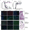
High titers of autoantibodies against glutamic acid decarboxylase (GAD) 65 are commonly observed in patients suffering from type 1 diabetes as well as stiff-person syndrome (SPS), a disorder that affects the CNS, and a variant of SPS, progressive encephalomyelitis with rigidity and myoclonus. Although there is a considerable amount of data focusing on the role of GAD65-specific CD4(+) T cells in type 1 diabetes, little is known about their role in SPS. In this study, we show that mice possessing a monoclonal GAD65-specific CD4(+) T cell population (4B5, PA19.9G11, or PA17.9G7) develop a lethal encephalomyelitis-like disease in the absence of any other T cells or B cells. GAD65-reactive CD4(+) T cells were found throughout the CNS in direct concordance with GAD65 expression and activated microglia: proximal to the circumventricular organs at the interface between the brain parenchyma and the blood-brain barrier. In the presence of B cells, high titer anti-GAD65 autoantibodies were generated, but these had no effect on the incidence or severity of disease. In addition, GAD65-specific CD4(+) T cells isolated from the brain were activated and produced IFN-gamma. These findings suggest that GAD65-reactive CD4(+) T cells alone mediate a lethal encephalomyelitis-like disease that may serve as a useful model to study GAD65-mediated diseases of the CNS.
Figures


Similar articles
-
CD4(+) T cells from glutamic acid decarboxylase (GAD)65-specific T cell receptor transgenic mice are not diabetogenic and can delay diabetes transfer.J Exp Med. 2002 Aug 19;196(4):481-92. doi: 10.1084/jem.20011845. J Exp Med. 2002. PMID: 12186840 Free PMC article.
-
Prevention of type I diabetes transfer by glutamic acid decarboxylase 65 peptide 206-220-specific T cells.Proc Natl Acad Sci U S A. 2004 Sep 28;101(39):14204-9. doi: 10.1073/pnas.0405500101. Epub 2004 Sep 20. Proc Natl Acad Sci U S A. 2004. PMID: 15381770 Free PMC article.
-
Glutamic acid decarboxylase T lymphocyte responses associated with susceptibility or resistance to type I diabetes: analysis in disease discordant human twins, non-obese diabetic mice and HLA-DQ transgenic mice.Int Immunol. 1998 Dec;10(12):1765-76. doi: 10.1093/intimm/10.12.1765. Int Immunol. 1998. PMID: 9885897
-
Immunobiology of stiff-person syndrome.Int Rev Immunol. 2008 Jan-Apr;27(1-2):79-92. doi: 10.1080/08830180701883240. Int Rev Immunol. 2008. PMID: 18300057 Review.
-
Stiff-person syndrome (SPS) and anti-GAD-related CNS degenerations: protean additions to the autoimmune central neuropathies.J Autoimmun. 2011 Sep;37(2):79-87. doi: 10.1016/j.jaut.2011.05.005. Epub 2011 Jun 16. J Autoimmun. 2011. PMID: 21680149 Review.
Cited by
-
Stiff-person syndrome and related disorders - diagnosis, mechanisms and therapies.Nat Rev Neurol. 2024 Oct;20(10):587-601. doi: 10.1038/s41582-024-01012-3. Epub 2024 Sep 3. Nat Rev Neurol. 2024. PMID: 39227464 Review.
-
Repositioning synthetic glucocorticoids in psychiatric disease associated with neural autoantibodies: a narrative review.J Neural Transm (Vienna). 2023 Aug;130(8):1029-1038. doi: 10.1007/s00702-022-02578-2. Epub 2022 Dec 28. J Neural Transm (Vienna). 2023. PMID: 36576564 Free PMC article. Review.
-
Orchestrated leukocyte recruitment to immune-privileged sites: absolute barriers versus educational gates.Nat Rev Immunol. 2013 Mar;13(3):206-18. doi: 10.1038/nri3391. Nat Rev Immunol. 2013. PMID: 23435332 Review.
-
GAD antibodies in neurological disorders - insights and challenges.Nat Rev Neurol. 2020 Jul;16(7):353-365. doi: 10.1038/s41582-020-0359-x. Epub 2020 May 26. Nat Rev Neurol. 2020. PMID: 32457440 Review.
-
Autoimmune stiff person syndrome and related myelopathies: understanding of electrophysiological and immunological processes.Muscle Nerve. 2012 May;45(5):623-34. doi: 10.1002/mus.23234. Muscle Nerve. 2012. PMID: 22499087 Free PMC article. Review.
References
-
- Tisch R, Yang XD, Singer SM, Liblau RS, Fugger L, McDevitt HO. Immune response to glutamic acid decarboxylase correlates with insulitis in non-obese diabetic mice. Nature. 1993;366:72–75. - PubMed
-
- Solimena M, Folli F, Aparisi R, Pozza G, De Camilli P. Autoantibodies to GABA-ergic neurons and pancreatic beta cells in stiff-man syndrome. The New England journal of medicine. 1990;322:1555–1560. - PubMed
-
- Solimena M, Folli F, Denis-Donini S, Comi GC, Pozza G, De Camilli P, Vicari AM. Autoantibodies to glutamic acid decarboxylase in a patient with stiff-man syndrome, epilepsy, and type I diabetes mellitus. The New England journal of medicine. 1988;318:1012–1020. - PubMed
-
- Baekkeskov S, Aanstoot HJ, Christgau S, Reetz A, Solimena M, Cascalho M, Folli F, Richter-Olesen H, De Camilli P. Identification of the 64K autoantigen in insulin-dependent diabetes as the GABA-synthesizing enzyme glutamic acid decarboxylase. Nature. 1990;347:151–156. - PubMed
Publication types
MeSH terms
Substances
Grants and funding
LinkOut - more resources
Full Text Sources
Other Literature Sources
Molecular Biology Databases
Research Materials

