. 2010 Mar;22(3):973-90.
doi: 10.1105/tpc.109.069658. Epub 2010 Mar 26.

Innate immune responses activated in Arabidopsis roots by microbe-associated molecular patterns

Affiliations

Innate immune responses activated in Arabidopsis roots by microbe-associated molecular patterns

Yves A Millet et al. Plant Cell. 2010 Mar.

Abstract

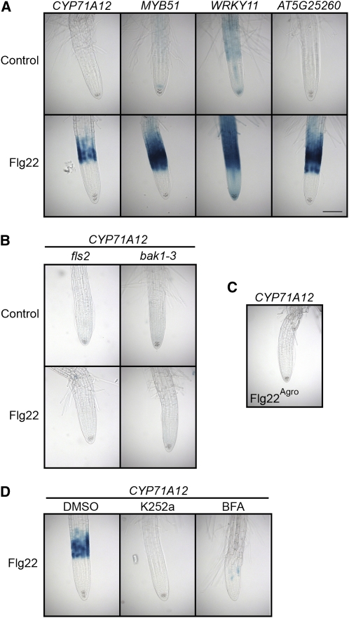
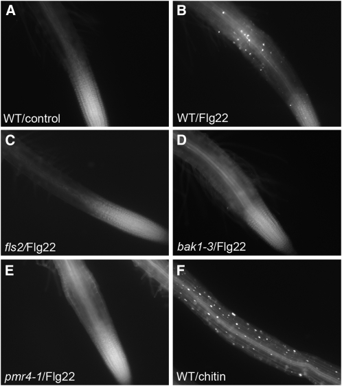
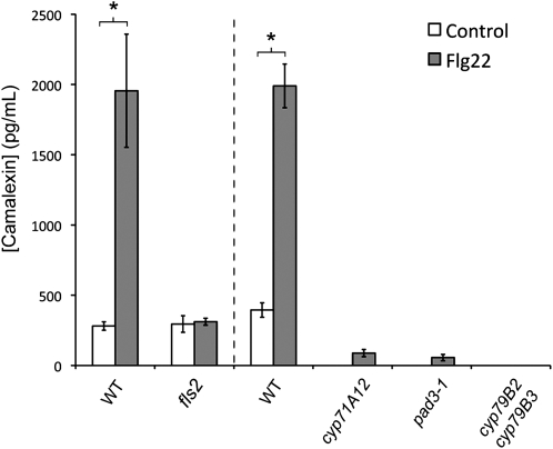
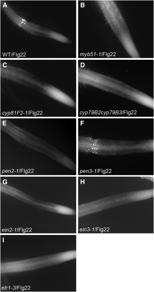
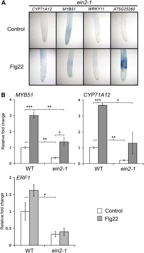
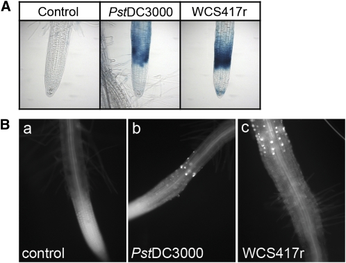
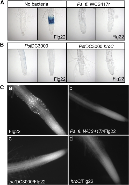
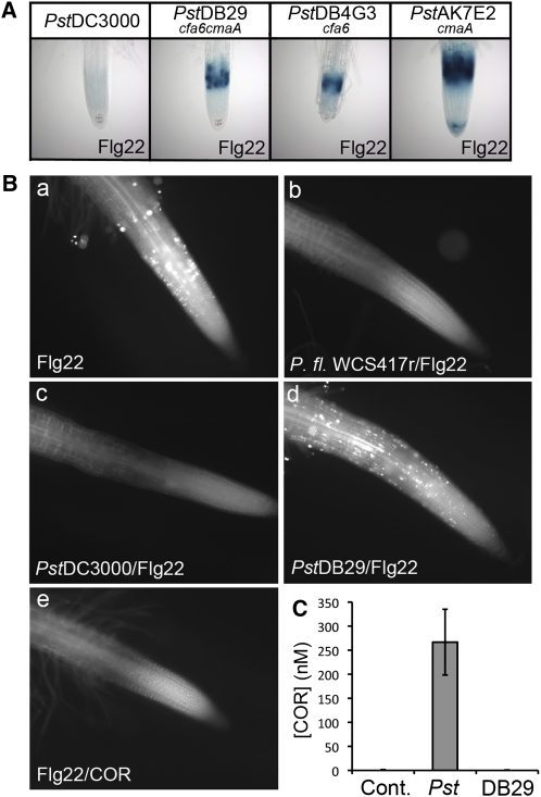
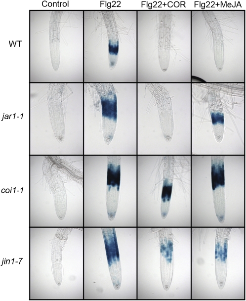
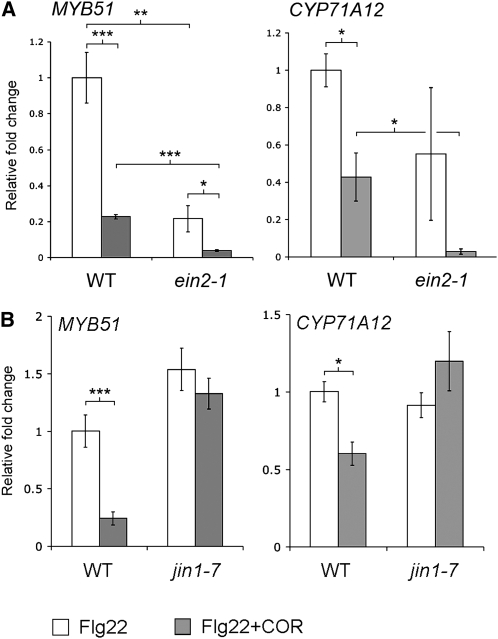
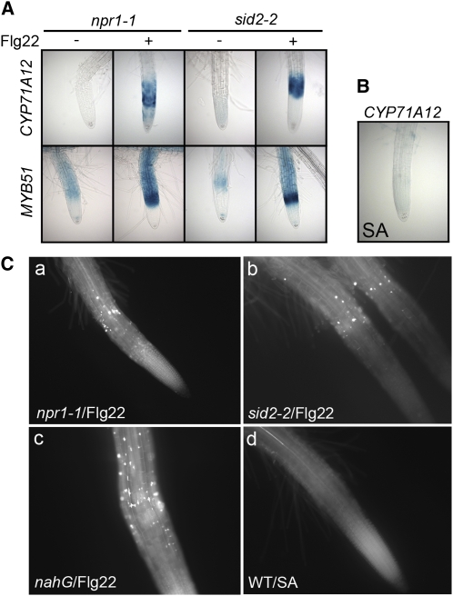
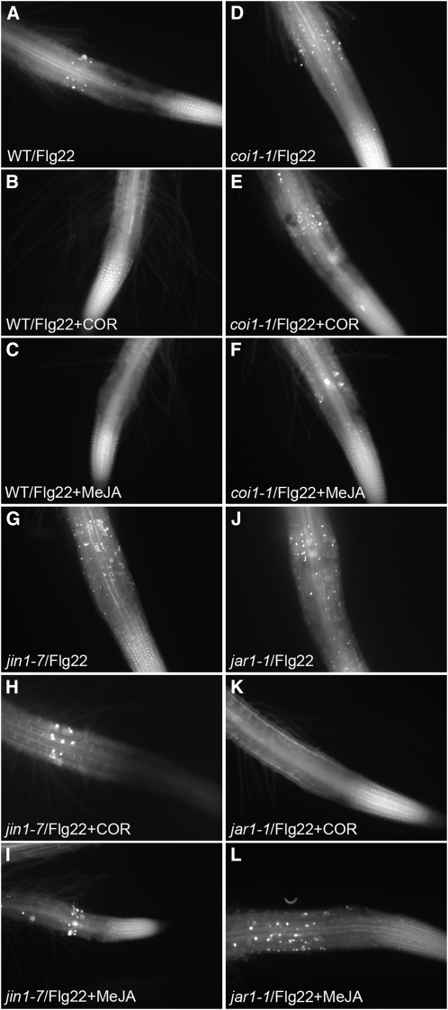
Despite the fact that roots are the organs most subject to microbial interactions, very little is known about the response of roots to microbe-associated molecular patterns (MAMPs). By monitoring transcriptional activation of beta-glucuronidase reporters and MAMP-elicited callose deposition, we show that three MAMPs, the flagellar peptide Flg22, peptidoglycan, and chitin, trigger a strong tissue-specific response in Arabidopsis thaliana roots, either at the elongation zone for Flg22 and peptidoglycan or in the mature parts of the roots for chitin. Ethylene signaling, the 4-methoxy-indole-3-ylmethylglucosinolate biosynthetic pathway, and the PEN2 myrosinase, but not salicylic acid or jasmonic acid signaling, play major roles in this MAMP response. We also show that Flg22 induces the cytochrome P450 CYP71A12-dependent exudation of the phytoalexin camalexin by Arabidopsis roots. The phytotoxin coronatine, an Ile-jasmonic acid mimic produced by Pseudomonas syringae pathovars, suppresses MAMP-activated responses in the roots. This suppression requires the E3 ubiquitin ligase COI1 as well as the transcription factor JIN1/MYC2 but does not rely on salicylic acid-jasmonic acid antagonism. These experiments demonstrate the presence of highly orchestrated and tissue-specific MAMP responses in roots and potential pathogen-encoded mechanisms to block these MAMP-elicited signaling pathways.


Figures

Figure 1.
Figure 1.
Flg22 Elicits Promoter:GUS Reporter Gene Expression in Transgenic Arabidopsis Seedlings. (A) Flg22 elicits expression of GUS reporter genes in the root EZ. Transgenic seedlings carrying CYP71A12pro:GUS, MYB51pro:GUS, WRKY11pro:GUS, or AT5G25260pro:GUS reporters were treated with 100 nM Flg22 or an equal volume of water as a control for 3 h (MYB51 and WRKY11) or 5 h (CYP71A12 and AT5G25260) before GUS staining. Bar = 100 μm. (B) Flg22 elicitation of CYP71A12pro:GUS depends on the Flg22 receptor FLS2 and the accessory receptor-like kinase BAK1. Transgenic fls2 CYP71A12pro:GUS or bak1-3 CYP71A12pro:GUS seedlings were treated with 100 nM Flg22 or water for 5 h before GUS staining. (C) A peptide corresponding to A. tumefaciens flagellin does not activate CYP71A12pro:GUS. Transgenic CYP71A12pro:GUS seedlings were treated with 100 nM Flg22Agro for 5 h before GUS staining. (D) Flg22 elicitation of a CYP71A12pro:GUS is blocked by the kinase inhibitor K252a and the membrane transport inhibitor BFA. Transgenic CYP71A12pro:GUS seedlings were cotreated with 100 nM Flg22 plus 1% DMSO, 1 μM K252a in DMSO, or 100 μg/mL BFA in DMSO for 5 h before GUS staining.
Figure 2.
Figure 2.
Flg22 and Chitin-Elicited Callose Deposition in Wild-Type and Mutant Arabidopsis Roots. Callose staining in roots of seedlings treated with water (A), 1 μM Flg22 ([B] to [E]), or 500 μg/mL chitin (F) for 18 h. Col-0 ([A],[B], and [F]); fls2 (C); bak1-3 (D); and pmr4-1 (E).
Figure 3.
Figure 3.
Flg22 Activates the Exudation of Camalexin in the Roots via CYP71A12. Liquid chromatography–mass spectrometry (LC-MS) analysis of camalexin in the exudate of 15-d-old seedling roots treated with 1 μM Flg22 for 24 h. Data represent the mean ± se of three replicate samples. *P < 0.05; two-tailed t test.
Figure 4.
Figure 4.
MYB51, CYP81F2, PEN2, and ET Signaling Are Required for Flg22-Elicited Callose Deposition in Roots. Callose staining in the roots of Col-0 (A); myb51-1 (B), cyp81F2-1 (C), cyp79B2 cyp79B3 (D), pen2-1 (E), pen3-1 (F), ein2-1 (G), ein3-1 (H), or etr1-3 (I) treated with 1 μM Flg22 for 18 h.
Figure 5.
Figure 5.
The Flg22 Response in the Roots Is ET Dependent. (A) Transgenic seedlings carrying CYP71A12pro:GUS, MYB51pro:GUS, or WRKY11pro:GUS reporters in an ein2-1 mutant background were treated with 100 nM Flg22 or water for 3 h (for MYB51 and WRKY11) or 5 h (for CYP71A12 and AT5G25260) before GUS staining. (B) qRT-PCR analysis of MYB51, CYP71A12, and ERF1 transcript levels in the roots of 2-week-old Col-0 or ein2-1 seedlings grown on vertical plates and treated with 1 μM Flg22 or water for 3 h. Data represent the mean ± sd of three replicates. *P < 0.05, **P < 0.01, ***P < 0.001; two-tailed t test.
Figure 6.
Figure 6.
Heat-Killed P. syringae DC3000 and P. fluorescens WCS417r Activate the CYP71A12pro:GUS Reporter and Callose Deposition. (A) Heat-killed Pst DC3000 and Ps. fl. WCS417r activate the CYP71A12pro:GUS reporter expression in the root EZ. Transgenic seedlings carrying CYP71A12pro:GUS were treated with heat-killed bacteria at a final OD600 of 0.1 or an equal volume of water as a control for 5 h before GUS staining. (B) Heat-killed Pst DC3000 and Ps. fl. WCS417r activate the deposition of callose in the EZ of Arabidopsis roots. Col-0 seedlings were treated with heat-killed bacteria at a final OD600 of 0.1 for 18 h before callose staining.
Figure 7.
Figure 7.
P. syringae and P. fluorescens Suppress Flg22-Elicited Responses in Arabidopsis Roots. (A) P. fluorescens WCS417r suppresses Flg22-elicited expression of CYP71A12pro:GUS. Transgenic CYP71A12pro:GUS seedlings were treated with 100 nM Flg22 for 5 h or preinfected at an initial OD600 of 0.002 with WCS417r for 18 h and then treated with 100 nM Flg22 for 5 h before GUS staining. (B) The P. syringae DC3000 type III secretion system is not required for suppression of Flg22-elicited expression of CYP71A12pro:GUS. Transgenic CYP71A12pro:GUS seedlings were preinfected at an initial OD600 of 0.002 with Pst DC3000 or Pst DC3000 hrcC (CUCPB5112) for 18 h and then treated with 100 nM Flg22 for 5 h before GUS staining. The final bacterial titers were ∼108 cells per mL for Pst DC3000 and Pst DC3000 hrcC and 109 cells per mL for Ps. fl. WCS417r (C) P. syringae DC3000 and P. fluorescens WCS417r suppress the Flg22-elicited deposition of callose in Arabidopsis roots. Col-0 seedlings treated with 1 μM Flg22 for 18 h (a) or preinfected with P. fl. WCS417r (b), Pst DC3000 (c), or Pst DC3000 CUCPB5112 (hrcC) (d) for 12 h and then treated with 1 μM Flg22 for 18 h.
Figure 8.
Figure 8.
COR Secreted by P. syringae DC3000 Suppresses the Flg22-Elicited Responses in Arabidopsis Roots. (A) COR synthesized by P. syringae DC3000 suppresses Flg22-elicited expression of CYP71A12pro:GUS. Col-0 seedlings media were infected at an initial OD600 of 0.002 with Pst DC3000 or the COR-deficient mutant DB29 (cfa;cma), DB4G3 (cfa), or AK7E2 (cma) for 22 h. The collected media (exudate) were filtered. Transgenic CYP71A12pro:GUS seedlings were incubated in the filtered media for 1 h and treated with 100 nM Flg22 for 5 h before GUS staining. The final bacterial titers were ∼108 cells per mL for all bacterial strains. (B) COR suppresses the Flg22-elicited deposition of callose in Arabidopsis roots. Col-0 seedlings were treated with 1 μM Flg22 for 18 h (a), pretreated with P. fluorescens WCS417r exudate (b), Pst DC3000 exudate (c), or Pst DB29 (cfa;cma) exudate (d) for 1 h and then treated with 1 μM Flg22 for 18 h, cotreated with 1 μM Flg22 and 1 μM COR for 18 h (e). (C) LC-MS analysis of the amount of COR secreted by Pst DC3000 and Pst DB29 in the media of Col-0 seedlings infected for 18 h. Data represent the mean ±sd of three replicates.
Figure 9.
Figure 9.
COR Acts as a Mimic of JA-Ile in Suppressing Flg22-Elicited Expression of CYP71A12pro:GUS in Arabidopsis Roots. Transgenic seedlings carrying a CYP71A12pro:GUS reporter construct in the wild type, jar1-1, coi1-1, or jin1-7 backgrounds were cotreated with 1 μM COR or 10 μM MeJA and 100 nM Flg22 or with an equal volume of water as a control for 5 h before GUS staining.
Figure 10.
Figure 10.
COR Suppresses Both the ET-Dependent and -Independent Flg22-Elicited Activation of MYB51 and CYP71A12 and Requires JIN1/MYC2 for Suppression. qRT-PCR analysis of MYB51 and CYP71A12 transcript levels in the roots of 2-week-old Col-0 and ein2-1 (A) or Col-0 and jin1-7 (B) seedlings grown on vertical plates and treated with 1 μM Flg22 with or without 0.2 μM COR for 3 h. Data represent the mean ± sd of three replicate samples. *P < 0.05, **P < 0.01, ***P < 0.001; two-tailed t test.
Figure 11.
Figure 11.
The Flg22-Elicited Response Suppressed by COR in Roots Is Independent of SA Signaling. (A) Flg22 elicited CYP71A12pro:GUS or MYB51pro:GUS expression in Arabidopsis seedlings. CYP71A12pro:GUS or MYB51pro:GUS seedlings were treated with 100 nM Flg22 for 3 h (for MYB51) or 5 h (for CYP71A12) in npr1-1 or sid2-2 mutant backgrounds. (B) CYP71A12pro:GUS seedlings were pretreated with 100 μM SA for 6, 12, or 24 h. No GUS staining was detected at any time point. (C) Callose deposition in the roots of Arabidopsis seedlings. npr1-1 (a), sid2-2 (b), nahG (c), or Col-0 (d) seedlings treated with 1 μM Flg22 for 18 h ([a] to [c]) or 100 μM SA for 18 h (d).
Figure 12.
Figure 12.
The COR-Mediated Suppression of the Flg22-Elicited Callose Deposition in Roots Requires COI1 and JIN1/MYC2. Callose staining in the roots of Col-0 ([A] to[C]), coi1-1 ([D] to[F]), jin1-7 ([G] to[I]); or jar1-1 ([J] to[L]) treated with 1 μM Flg22 ([A], [D],[G], and[J]), 1 μM Flg22 and 1 μM COR ([B],[E],[H], and [K]), or 1 μM Flg22 and 10 μM MeJA ([C],[F],[I], and [L]) for 18 h.

Similar articles

Cited by

References

    1. Aist J.R., Bushnell W.R. (1991). Invasion of plants by powdery mildew fungi, and cellular mechanisms of resistance. In The Fungal Spore and Disease Initiation in Plants and Animals, Cole G.T., Mock H.C., (New York: Plenum; ), pp. 321–345
    1. Andreote F.D., de Araujo W.L., de Azevedo J.L., van Elsas J.D., da Rocha U.N., van Overbeek L.S. (2009). Endophytic colonization of potato (Solanum tuberosum L.) by a novel competent bacterial endophyte, Pseudomonas putida strain P9, and its effect on associated bacterial communities. Appl. Environ. Microbiol. 75: 3396–3406 - PMC - PubMed
    1. Attard A., Gourgues M., Galiana E., Panabieres F., Ponchet M., Keller H. (2008). Strategies of attack and defense in plant-oomycete interactions, accentuated for Phytophthora parasitica Dastur (syn. P. Nicotianae Breda de Haan). J. Plant Physiol. 165: 83–94 - PubMed
    1. Badri D.V., Vivanco J.M. (2009). Regulation and function of root exudates. Plant Cell Environ. 32: 666–681 - PubMed
    1. Bais H.P., Fall R., Vivanco J.M. (2004). Biocontrol of Bacillus subtilis against infection of Arabidopsis roots by Pseudomonas syringae is facilitated by biofilm formation and surfactin production. Plant Physiol. 134: 307–319 - PMC - PubMed

Publication types

MeSH terms

LinkOut - more resources