. 2010 Mar;22(3):729-43.
doi: 10.1105/tpc.109.068734. Epub 2010 Mar 26.

The Arabidopsis stem cell factor POLTERGEIST is membrane localized and phospholipid stimulated

Affiliations

The Arabidopsis stem cell factor POLTERGEIST is membrane localized and phospholipid stimulated

Jennifer M Gagne et al. Plant Cell. 2010 Mar.

Abstract

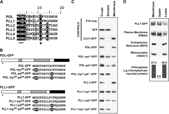
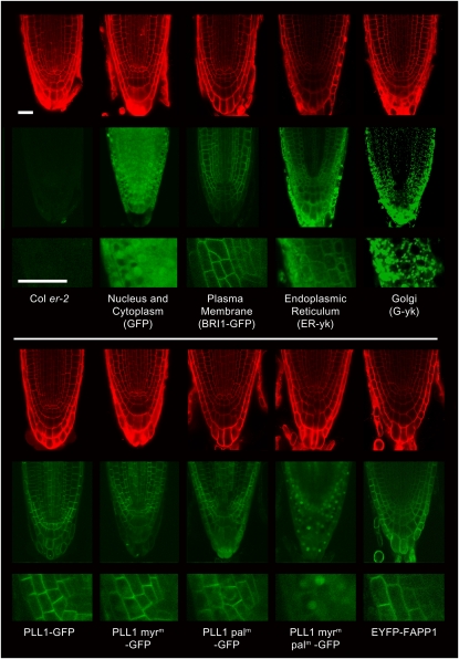
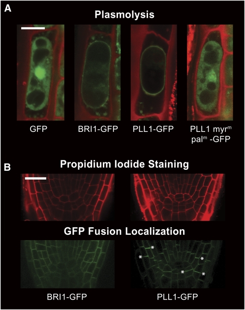
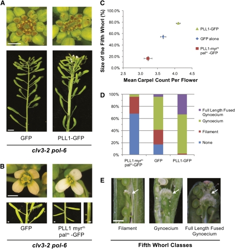
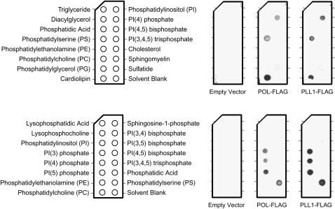
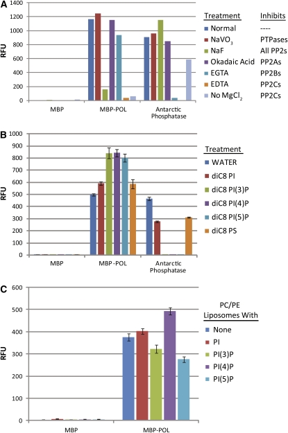
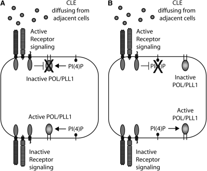
Stem cell maintenance and differentiation are tightly regulated in multicellular organisms. In plants, proper control of the stem cell populations is critical for extensive postembryonic organogenesis. The Arabidopsis thaliana protein phosphatase type 2C proteins POLTERGEIST (POL) and PLL1 are essential for maintenance of both the root and shoot stem cells. Specifically, POL and PLL1 are required for proper specification of key asymmetric cell divisions during stem cell initiation and maintenance. POL and PLL1 are known to be integral components of the CLE/WOX signaling pathways, but the location and mechanisms by which POL and PLL1 are regulated within these pathways are unclear. Here, we show that POL and PLL1 are dual-acylated plasma membrane proteins whose membrane localization is required for proper function. Furthermore, this localization places POL and PLL1 in proximity of the upstream plasma membrane receptors that regulate their activity. Additionally, we find that POL and PLL1 directly bind to multiple lipids and that POL is catalytically activated by phosphatidylinositol (4) phosphate [PI(4)P] in vitro. Based on these results, we propose that the upstream receptors in the CLE/WOX signaling pathways may function to either limit PI(4)P availability or antagonize PI(4)P stimulation of POL/PLL1. Significantly, the findings presented here suggest that phospholipids play an important role in promoting stem cell specification.


Figures

Figure 1.
Figure 1.
N-Myristoylation and -Palmitoylation Are Required for Plasma Membrane Localization of POL and PLL1. (A) Alignment of putative N-myristoylation and -palmitoylation signal peptides of the Arabidopsis POL family members. Positions with >66% amino acid similarity or conservation are indicated. The conserved and similar amino acids are in black and gray boxes, respectively. Residues mutated in this study are underlined. The asterisk marks the conserved Cys residues. (B) Structure of fusion proteins encoded by the 35S:POL-GFP and 35S:PLL1-GFP constructs. GFP is in black, while gray designates the regions with homology to the human PP2Ca catalytic domain. The asterisk marks the putative signal peptides. Below the structures are the first 18 amino acids for each of the constructs used with the amino acid substitutions marked by black boxes. (C) Immunoblots of crude, soluble, and membrane protein fractions from infiltrated tobacco plants expressing various GFP fusion proteins. (D) Two-phase partitioning of membranes isolated from infiltrated tobacco plants expressing PLL1-GFP. Various antibodies were used to show the partitioning of PLL1-GFP, the plasma membrane, the ER, and the mitochondrial membrane. Chlorophyll levels were also quantified for each fraction and are expressed in micrograms of chlorophyll per milligram of protein for each sample.
Figure 2.
Figure 2.
Myristoylation and Palmitoylation Are Required for Plasma Membrane Localization of PLL1 in Arabidopsis. Images are of roots of 4-d-old transgenic Arabidopsis expressing various fusion proteins and controls. The top images show the propidium iodide–stained cell walls (red), while the middle images show the localization patterns of the GFP (or YFP) fusion protein (green). The bottom images are enlargements of the same regions (bottom left of the middle images) from each of the GFP (or YFP) images to more clearly show the localization patterns. Bars = 25 μm.
Figure 3.
Figure 3.
PLL1 Is Irregularly Distributed in the Plasma Membrane of Arabidopsis Root Cells. (A) Plasmolysis of Arabidopsis root epidermal cells expressing various GFP fusion proteins. Images are an overlay of propidium iodide staining showing cell outlines in red with the localization of the GFP fusion proteins in green. Images were taken of 4-d-old seedlings following 2 h treatment with 0.8 M mannitol. Bar = 5 μm. (B) Images of roots of 4-d-old transgenic Arabidopsis expressing BRI1-GFP or PLL1-GFP taken using decreased sensitivity of the photomultiplier detector tube. The top images are of propidium iodide staining, while the bottom images show localization of the GFP fusion proteins. A few of the cell faces showing increased accumulation of PLL1-GFP are marked with asterisks. Bar = 25 μm.
Figure 4.
Figure 4.
Plasma Membrane Localization Is Required for Proper PLL1 Function in Planta. (A) and (B) Phenotypic analysis of pol-6 clv3-2 double mutants transformed with GFP, PLL1-GFP, or PLL1-myrmpalm-GFP. Images are of the shoot meristems and primary inflorescences (A) (bars = 5 mm) and representative flowers, siliques, and pedicles (B) (bars = 1 mm) of T1 plants. (C) Relative size of the fifth whorl and the mean number of carpels per flower for clv3-2 pol-6 plants transformed with GFP, PLL1-GFP, or PLL1-myrmpalm-GFP (n = 70, 50, and 50, respectively). Measurements and carpel counts are the average of the first 10 flowers from the primary inflorescences of 32-d-old T2 plants. The measured size of the fifth whorl is represented as a percentage of total silique length. The size of the silique is defined as the length between the attachment site for the sepals, petals, and stamens and the top of the gynoecium. The same definition was used to measure the fifth-whorl length. The vertical and horizontal bars represent se of the mean for the relative fifth-whorl size and the number of carpels, respectively. (D) Graph showing the phenotypic class distribution of the fifth whorls for the flowers analyzed in (C). (E) Representative images of the three different classes of fifth whorls (bar = 0.5 mm). Arrows are used to denote the fifth whorls.
Figure 5.
Figure 5.
POL and PLL1 Bind Phospholipids. POL-FLAG and PLL1-FLAG proteins were used to probe membranes spotted with the indicated lipids, and the fusion proteins were then detected with anti-FLAG antibody. Purified extract from an empty vector preparation was also tested as a negative control.
Figure 6.
Figure 6.
POL Activity Is Stimulated by PI(4)P. (A) MBP-POL phosphatase activity is specifically inhibited by PP2C phosphatase inhibitors and not by inhibitors that affect other classes of phosphatases. MBP and antarctic phosphatase are included as controls. (B) The effects of various water-soluble diC8 lipids on MBP-POL phosphatase activity. Antarctic phosphatase and MBP are included as controls. Data represent the average of three replicates, and the sd is shown. (C) The effects of PC/PE liposomes, containing no additional lipids, 5% PI, 5% PI(3)P, 5% PI(4)P, or 5% PI(5)P on MBP-POL phosphatase activity. Data represent the average of three replicates, and the sd is shown.
Figure 7.
Figure 7.
Two Potential Models for How CLE Signaling and PI(4)P Binding Regulate POL and PLL1 Activity in Planta. (A) In the first model, the two regulatory signals work antagonistically to set up a polar distribution of POL and PLL1 activity within the cell prior to cell division. (B) In the second model, CLE signaling negatively regulates PI(4)P levels on the apical side of the cell leading to a polar distribution of POL and PLL1 activity.

Similar articles

Cited by

References

    1. Alvarez-Venegas R., Sadder M., Hlavacka A., Baluska F., Xia Y., Lu G., Firsov A., Sarath G., Moriyama H., Dubrovsky J.G., Avramova Z. (2006). The Arabidopsis homolog of trithorax, ATX1, binds phosphatidylinositol 5-phosphate, and the two regulate a common set of target genes. Proc. Natl. Acad. Sci. USA 103: 6049–6054 - PMC - PubMed
    1. Batistic O., Sorek N., Schültke S., Yalovsky S., Kudla J. (2008). Dual fatty acyl modification determines the localization and plasma membrane targeting of CBL/CIPK Ca2+ signaling complexes in Arabidopsis. Plant Cell 20: 1346–1362 - PMC - PubMed
    1. Baudouin E., Meskiene I., Hirt H. (1999). Short communication: Unsaturated fatty acids inhibit MP2C, a protein phosphatase 2C involved in the wound-induced MAP kinase pathway regulation. Plant J. 20: 343–348 - PubMed
    1. Boisson B., Giglione C., Meinnel T. (2003). Unexpected protein families including cell defense components feature in the N-myristoylome of a higher eukaryote. J. Biol. Chem. 278: 43418–43429 - PubMed
    1. Brand U., Fletcher J.C., Hobe M., Meyerowitz E.M., Simon R. (2000). Dependence of stem cell fate in Arabidopsis on a feedback loop regulated by CLV3 activity. Science 289: 617–619 - PubMed

Publication types

MeSH terms