

Human CD8(+) T cells clear Cryptosporidium parvum from infected intestinal epithelial cells
- PMID: 20348507
- PMCID: PMC2844566
- DOI: 10.4269/ajtmh.2010.09-0590
Human CD8(+) T cells clear Cryptosporidium parvum from infected intestinal epithelial cells
Abstract
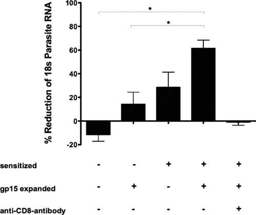
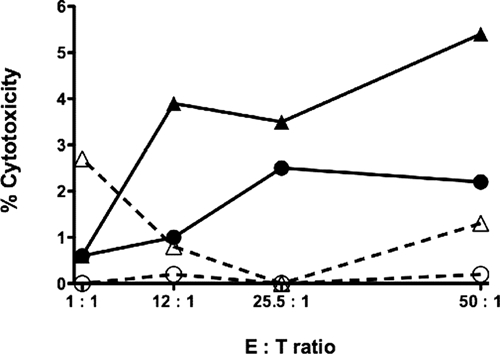
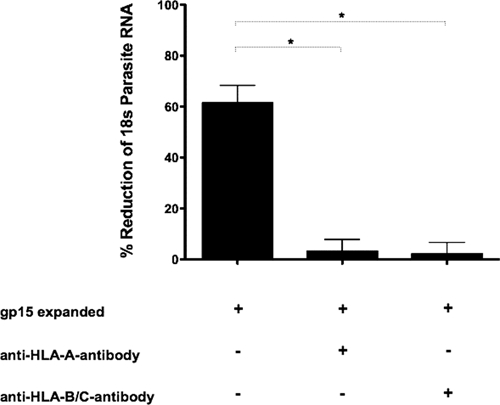
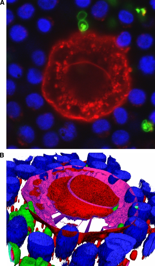
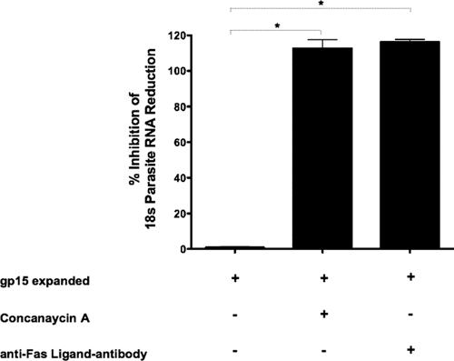
Intracellular protozoans of the genus Cryptosporidium are a major cause of diarrheal illness worldwide, especially in immunocompromised individuals. CD4(+) T cells and interferon-gamma are key factors in the control of cryptosporidiosis in human and murine models. Previous studies led us to hypothesize that CD8(+) T cells contribute to clearance of intestinal epithelial Cryptosporidium infection in humans. We report here that antigen expanded sensitized CD8(+) T cells reduce the parasite load in infected intestinal epithelial cell cultures and lyse infected intestinal epithelial cells. These effects are most likely mediated by the release of cytotoxic granules. Elimination of parasites seems to require antigen presentation through both human leukocyte antigen (HLA)-A and HLA-B. These data suggest that cytotoxic CD8(+) T cells play a role in clearing Cryptosporidium from the intestine, a previously unrecognized feature of the human immune response against this parasite.
Figures
Similar articles
-
Interleukin-15 activates human natural killer cells to clear the intestinal protozoan cryptosporidium.J Infect Dis. 2005 Oct 1;192(7):1294-302. doi: 10.1086/444393. Epub 2005 Aug 23. J Infect Dis. 2005. PMID: 16136475
-
Neonatal Mouse Gut Metabolites Influence Cryptosporidium parvum Infection in Intestinal Epithelial Cells.mBio. 2020 Dec 15;11(6):e02582-20. doi: 10.1128/mBio.02582-20. mBio. 2020. PMID: 33323514 Free PMC article.
-
Attenuation of Intestinal Epithelial Cell Migration During Cryptosporidium parvum Infection Involves Parasite Cdg7_FLc_1030 RNA-Mediated Induction and Release of Dickkopf-1.J Infect Dis. 2018 Sep 8;218(8):1336-1347. doi: 10.1093/infdis/jiy299. J Infect Dis. 2018. PMID: 30052999 Free PMC article.
-
Innate immune responses play a key role in controlling infection of the intestinal epithelium by Cryptosporidium.Int J Parasitol. 2017 Oct;47(12):711-721. doi: 10.1016/j.ijpara.2017.08.001. Epub 2017 Sep 8. Int J Parasitol. 2017. PMID: 28893638 Review.
-
Bovine T cell responses to Cryptosporidium parvum infection.Int J Parasitol. 1998 Jul;28(7):1083-8. doi: 10.1016/s0020-7519(98)00077-0. Int J Parasitol. 1998. PMID: 9724879 Review.
Cited by
-
Systemic and Mucosal Immune Responses to Cryptosporidium-Vaccine Development.Curr Trop Med Rep. 2015 Sep 1;2(3):171-180. doi: 10.1007/s40475-015-0054-y. Curr Trop Med Rep. 2015. PMID: 26279971 Free PMC article.
-
A review of the global burden, novel diagnostics, therapeutics, and vaccine targets for cryptosporidium.Lancet Infect Dis. 2015 Jan;15(1):85-94. doi: 10.1016/S1473-3099(14)70772-8. Epub 2014 Sep 29. Lancet Infect Dis. 2015. PMID: 25278220 Free PMC article. Review.
-
Structural Analyses of a Dominant Cryptosporidium parvum Epitope Presented by H-2Kb Offer New Options To Combat Cryptosporidiosis.mBio. 2023 Feb 28;14(1):e0266622. doi: 10.1128/mbio.02666-22. Epub 2023 Jan 5. mBio. 2023. PMID: 36602309 Free PMC article.
-
CD4+ T cells are not essential for control of early acute Cryptosporidium parvum infection in neonatal mice.Infect Immun. 2011 Apr;79(4):1647-53. doi: 10.1128/IAI.00922-10. Epub 2011 Jan 31. Infect Immun. 2011. PMID: 21282414 Free PMC article.
-
Immunity to Cryptosporidium: insights into principles of enteric responses to infection.Nat Rev Immunol. 2024 Feb;24(2):142-155. doi: 10.1038/s41577-023-00932-3. Epub 2023 Sep 11. Nat Rev Immunol. 2024. PMID: 37697084 Free PMC article. Review.
References
-
- Blanshard C, Jackson AM, Shanson DC, Francis N, Gazzard BG. Cryptosporidiosis in HIV-seropositive patients. Q J Med. 1992;85:813–823. - PubMed
-
- Flanigan T, Whalen C, Turner J, Soave R, Toerner J, Havlir D, Kotler D. Cryptosporidium infection and CD4 counts. Ann Intern Med. 1992;116:840–842. - PubMed
-
- Manabe YC, Clark DP, Moore RD, Lumadue JA, Dahlman HR, Belitsos PC, Chaisson RE, Sears CL. Cryptosporidiosis in patients with AIDS: correlates of disease and survival. Clin Infect Dis. 1998;27:536–542. - PubMed
-
- Dann SM, Wang HC, Gambarin KJ, Actor JK, Robinson P, Lewis DE, Caillat-Zucman S, White AC., Jr Interleukin-15 activates human natural killer cells to clear the intestinal protozoan Cryptosporidium. J Infect Dis. 2005;192:1294–1302. - PubMed
MeSH terms
Substances
Grants and funding
LinkOut - more resources
Full Text Sources
Research Materials
