

Proteomics analysis of the estrogen receptor alpha receptosome
- PMID: 20348541
- PMCID: PMC2938095
- DOI: 10.1074/mcp.M900457-MCP200
Proteomics analysis of the estrogen receptor alpha receptosome
Abstract
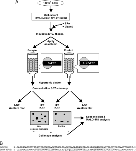
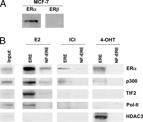
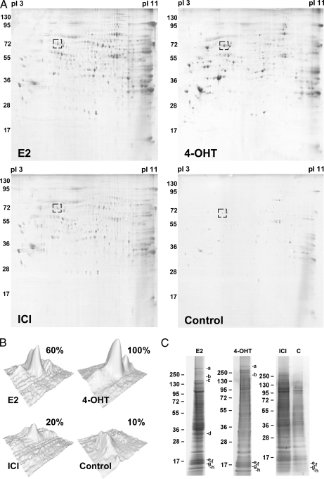
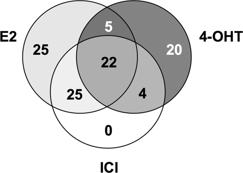
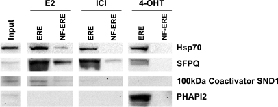
The estrogen receptors (ERs) are ligand-dependent transcription factors that activate transcription by binding to estrogen response elements. Estrogen-mediated effects are tissue- and cell type-specific, determined by the cofactor recruitment to the ERs among other factors. To understand these differences in estrogen action, it is important to identify the various compositions of the ER complexes (ER receptosomes). In this report, we describe a fast and efficient method for the isolation of the ERalpha receptosome for proteomics analysis. Using immobilized estrogen response element on a Sepharose column in combination with two-dimensional electrophoresis and MALDI-TOF MS, significant amounts of proteins could be isolated and identified. Differences in ERalpha complex composition with the ER ligands 17beta-estradiol, 4-hydroxytamoxifen, and ICI-182,780 could also be observed. Thus, this approach provides an easy and relevant way of identifying ERalpha cofactor and transcription factor recruitment under different conditions.
Figures
Similar articles
-
Exploration of dimensions of estrogen potency: parsing ligand binding and coactivator binding affinities.J Biol Chem. 2011 Apr 15;286(15):12971-82. doi: 10.1074/jbc.M110.205112. Epub 2011 Feb 14. J Biol Chem. 2011. PMID: 21321128 Free PMC article.
-
Structure activity relationships and differential interactions and functional activity of various equine estrogens mediated via estrogen receptors (ERs) ERalpha and ERbeta.Endocrinology. 2008 Oct;149(10):4857-70. doi: 10.1210/en.2008-0304. Epub 2008 Jul 3. Endocrinology. 2008. PMID: 18599548
-
Estrogen response element-dependent regulation of transcriptional activation of estrogen receptors alpha and beta by coactivators and corepressors.J Mol Endocrinol. 2004 Oct;33(2):387-410. doi: 10.1677/jme.1.01541. J Mol Endocrinol. 2004. PMID: 15525597
-
Rapid endocrine disruption: environmental estrogen actions triggered outside the nucleus.J Steroid Biochem Mol Biol. 2006 Dec;102(1-5):163-9. doi: 10.1016/j.jsbmb.2006.09.019. Epub 2006 Nov 3. J Steroid Biochem Mol Biol. 2006. PMID: 17084624 Review.
-
Estrogen receptor α and β in the normal immune system and in lymphoid malignancies.Mol Cell Endocrinol. 2013 Aug 15;375(1-2):121-9. doi: 10.1016/j.mce.2013.05.016. Epub 2013 May 23. Mol Cell Endocrinol. 2013. PMID: 23707618 Review.
Cited by
-
Age-dependent Effects of 17β-estradiol on the dynamics of estrogen receptor β (ERβ) protein-protein interactions in the ventral hippocampus.Mol Cell Proteomics. 2014 Mar;13(3):760-79. doi: 10.1074/mcp.M113.031559. Epub 2014 Jan 5. Mol Cell Proteomics. 2014. PMID: 24390426 Free PMC article.
-
Differential recruitment of co-regulatory proteins to the human estrogen receptor 1 in response to xenoestrogens.Comp Biochem Physiol Part D Genomics Proteomics. 2016 Sep;19:159-173. doi: 10.1016/j.cbd.2016.04.003. Epub 2016 Apr 20. Comp Biochem Physiol Part D Genomics Proteomics. 2016. PMID: 27156127 Free PMC article.
-
Identification of estrogen receptor proteins in breast cancer cells using matrix-assisted laser desorption/ionization time of flight mass spectrometry (Review).Oncol Lett. 2014 May;7(5):1341-1344. doi: 10.3892/ol.2014.1912. Epub 2014 Feb 26. Oncol Lett. 2014. PMID: 24765135 Free PMC article.
-
A meta-analysis to evaluate the cellular processes regulated by the interactome of endogenous and over-expressed estrogen receptor alpha.Oncoscience. 2015 Mar 7;2(5):487-496. doi: 10.18632/oncoscience.138. eCollection 2015. Oncoscience. 2015. PMID: 26097882 Free PMC article.
-
The Effects of 17β-estradiol in Cancer are Mediated by Estrogen Receptor Signaling at the Plasma Membrane.Front Physiol. 2011 Jun 30;2:30. doi: 10.3389/fphys.2011.00030. eCollection 2011. Front Physiol. 2011. PMID: 21747767 Free PMC article.
References
-
- Nilsson S., Gustafsson J. A. (2002) Estrogen receptor action. Crit. Rev. Eukaryot. Gene. Expr. 12, 237–257 - PubMed
-
- Behl C. (2002) Estrogen can protect neurons: modes of action. J. Steroid. Biochem. Mol. Biol. 83, 195–197 - PubMed
-
- Farhat M. Y., Lavigne M. C., Ramwell P. W. (1996) The vascular protective effects of estrogen. FASEB J. 10, 615–624 - PubMed
-
- Nilsson S., Mäkelä S., Treuter E., Tujague M., Thomsen J., Andersson G., Enmark E., Pettersson K., Warner M., Gustafsson J. A. (2001) Mechanisms of estrogen action. Physiol. Rev. 81, 1535–1565 - PubMed
-
- Matthews J., Gustafsson J. A. (2003) Estrogen signaling: a subtle balance between ER alpha and ER beta. Mol. Interv. 3, 281–292 - PubMed
Publication types
MeSH terms
Substances
LinkOut - more resources
Full Text Sources
