T helper type 1 and 17 cells determine efficacy of interferon-beta in multiple sclerosis and experimental encephalomyelitis
- PMID: 20348925
- PMCID: PMC3042885
- DOI: 10.1038/nm.2110
T helper type 1 and 17 cells determine efficacy of interferon-beta in multiple sclerosis and experimental encephalomyelitis
Abstract
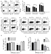
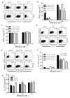
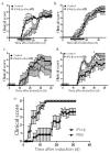
Interferon-beta (IFN-beta) is the major treatment for multiple sclerosis. However, this treatment is not always effective. Here we have found congruence in outcome between responses to IFN-beta in experimental autoimmune encephalomyelitis (EAE) and relapsing-remitting multiple sclerosis (RRMS). IFN-beta was effective in reducing EAE symptoms induced by T helper type 1 (T(H)1) cells but exacerbated disease induced by T(H)17 cells. Effective treatment in T(H)1-induced EAE correlated with increased interleukin-10 (IL-10) production by splenocytes. In T(H)17-induced disease, the amount of IL-10 was unaltered by treatment, although, unexpectedly, IFN-beta treatment still reduced IL-17 production without benefit. Both inhibition of IL-17 and induction of IL-10 depended on IFN-gamma. In the absence of IFN-gamma signaling, IFN-beta therapy was ineffective in EAE. In RRMS patients, IFN-beta nonresponders had higher IL-17F concentrations in serum compared to responders. Nonresponders had worse disease with more steroid usage and more relapses than did responders. Hence, IFN-beta is proinflammatory in T(H)17-induced EAE. Moreover, a high IL-17F concentration in the serum of people with RRMS is associated with nonresponsiveness to therapy with IFN-beta.
Figures


Comment in
-
Molecular oracles for multiple sclerosis therapy.Nat Med. 2010 Apr;16(4):376-7. doi: 10.1038/nm0410-376. Nat Med. 2010. PMID: 20376043 No abstract available.
Similar articles
-
IL-27 mediates the response to IFN-β therapy in multiple sclerosis patients by inhibiting Th17 cells.Brain Behav Immun. 2011 Aug;25(6):1170-81. doi: 10.1016/j.bbi.2011.03.007. Epub 2011 Mar 21. Brain Behav Immun. 2011. PMID: 21420486
-
Preferential recruitment of interferon-gamma-expressing TH17 cells in multiple sclerosis.Ann Neurol. 2009 Sep;66(3):390-402. doi: 10.1002/ana.21748. Ann Neurol. 2009. PMID: 19810097
-
Ethanol extract of Glycyrrhizae Radix modulates the responses of antigen-specific splenocytes in experimental autoimmune encephalomyelitis.Phytomedicine. 2019 Feb 15;54:56-65. doi: 10.1016/j.phymed.2018.09.189. Epub 2018 Sep 18. Phytomedicine. 2019. PMID: 30668383
-
Current views on the roles of Th1 and Th17 cells in experimental autoimmune encephalomyelitis.J Neuroimmune Pharmacol. 2010 Jun;5(2):189-97. doi: 10.1007/s11481-009-9188-9. Epub 2010 Jan 27. J Neuroimmune Pharmacol. 2010. PMID: 20107924 Free PMC article. Review.
-
Janus-like effects of type I interferon in autoimmune diseases.Immunol Rev. 2012 Jul;248(1):23-35. doi: 10.1111/j.1600-065X.2012.01131.x. Immunol Rev. 2012. PMID: 22725952 Free PMC article. Review.
Cited by
-
Type I interferons: beneficial in Th1 and detrimental in Th17 autoimmunity.Clin Rev Allergy Immunol. 2013 Apr;44(2):114-20. doi: 10.1007/s12016-011-8296-5. Clin Rev Allergy Immunol. 2013. PMID: 22231516 Free PMC article. Review.
-
Immune checkpoints in central nervous system autoimmunity.Immunol Rev. 2012 Jul;248(1):122-39. doi: 10.1111/j.1600-065X.2012.01136.x. Immunol Rev. 2012. PMID: 22725958 Free PMC article. Review.
-
Transplantation of human adipose-derived stem cells overexpressing LIF/IFN-β promotes recovery in experimental autoimmune encephalomyelitis (EAE).Sci Rep. 2022 Oct 25;12(1):17835. doi: 10.1038/s41598-022-21850-9. Sci Rep. 2022. PMID: 36284106 Free PMC article.
-
Type I interferons and microbial metabolites of tryptophan modulate astrocyte activity and central nervous system inflammation via the aryl hydrocarbon receptor.Nat Med. 2016 Jun;22(6):586-97. doi: 10.1038/nm.4106. Epub 2016 May 9. Nat Med. 2016. PMID: 27158906 Free PMC article.
-
The role of interferon-β in the treatment of multiple sclerosis and experimental autoimmune encephalomyelitis - in the perspective of inflammasomes.Immunology. 2013 May;139(1):11-8. doi: 10.1111/imm.12081. Immunology. 2013. PMID: 23360426 Free PMC article. Review.
References
-
- Arnason BG. Immunologic therapy of multiple sclerosis. Annu Rev Med. 1999;50:291–302. - PubMed
-
- Wang AG, Lin YC, Wang SJ, Tsai CP, Yen MY. Early relapse in multiple sclerosis-associated optic neuritis following the use of interferon beta-1a in Chinese patients. Jpn J Ophthalmol. 2006;50:537–42. - PubMed
-
- Benveniste EN, Qin H. Type I interferons as anti-inflammatory mediators. Sci STKE. 2007;2007:pe70. - PubMed
-
- Prinz M, et al. Distinct and Nonredundant In Vivo Functions of IFNAR on Myeloid Cells Limit Autoimmunity in the Central Nervous System. Immunity. 2008 - PubMed
Publication types
MeSH terms
Substances
Grants and funding
LinkOut - more resources
Full Text Sources
Other Literature Sources
Molecular Biology Databases

