. 2010 Jun 10;29(23):3398-410.
doi: 10.1038/onc.2010.94. Epub 2010 Mar 29.

The proapoptotic BH3-only protein Bim is downregulated in a subset of colorectal cancers and is repressed by antiapoptotic COX-2/PGE(2) signalling in colorectal adenoma cells

Affiliations

The proapoptotic BH3-only protein Bim is downregulated in a subset of colorectal cancers and is repressed by antiapoptotic COX-2/PGE(2) signalling in colorectal adenoma cells

A Greenhough et al. Oncogene. .

Abstract

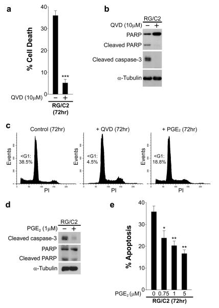
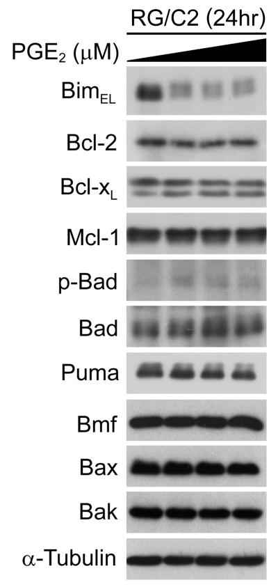
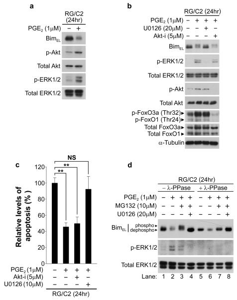
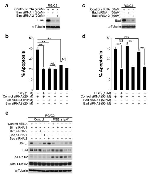
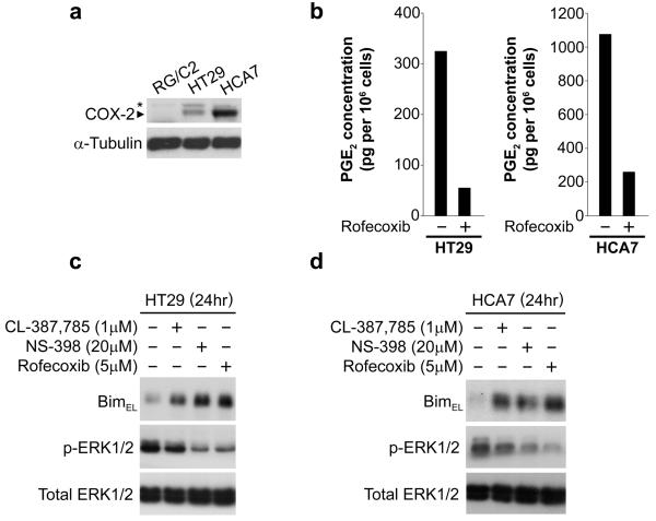
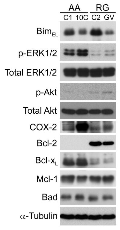
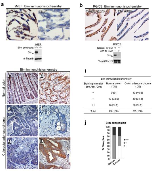
Overexpression of cyclooxygenase-2 (COX-2) and elevated levels of its enzymatic product prostaglandin E2 (PGE(2)) occur in the majority of colorectal cancers and have important roles in colorectal tumorigenesis. However, despite the established prosurvival role of PGE(2) in cancer, the underlying mechanisms are not fully understood. Here, we have shown that PGE(2) suppresses apoptosis via repression of the proapoptotic BH3-only protein Bim in human colorectal adenoma cells. Repression of Bim expression was dependent upon PGE(2)-mediated activation of the Raf-MEK-ERK1/2 pathway, which promoted Bim phosphorylation and proteasomal degradation. Reduction of Bim expression using RNA interference reduced spontaneous apoptosis in adenoma cells and abrogated PGE(2)-dependent apoptosis suppression. Treatment of COX-2-expressing colorectal carcinoma cells with COX-2-selective NSAIDs-induced Bim expression, suggesting that Bim repression via PGE(2) signalling may be opposed by COX-2 inhibition. Examination of Bim expression in two established in vitro models of the adenoma-carcinoma sequence revealed that downregulation of Bim expression was associated with tumour progression towards an anchorage-independent phenotype. Finally, immunohistochemical analyses revealed that Bim expression is markedly reduced in approximately 40% of human colorectal carcinomas in vivo. These observations highlight the COX-2/PGE(2) pathway as an important negative regulator of Bim expression in colorectal tumours and suggest that Bim repression may be an important step during colorectal cancer tumorigenesis.


Figures

Figure 1
Figure 1. PGE2 suppresses apoptosis in RG/C2 cells
(a) RG/C2 cells exhibit relatively high levels of spontaneous cell death that can be abolished by caspase inhibition. RG/C2 cells were cultured in the presence and absence of the broad spectrum caspase inhibitor QVD for 72 hours; the proportion of apoptotic cells was significantly reduced by QVD. Columns show the mean of three independent experiments performed in triplicate; ***p<0.001. (b) QVD prevents spontaneous caspase-3 and PARP cleavage in RG/C2 cells. Western blotting was performed on whole cell lysates from the total population of cells (attached plus floating). QVD increased intact PARP (116kDa) levels and inhibited PARP cleavage (89kDa). (c) QVD and PGE2 reduced the proportion of sub-G1 DNA-containing RG/C2 cells. Cells were treated with vehicle control, QVD, or PGE2, for 72 hours, after which the total population of cells was subjected to propidium iodide staining and flow cytometric analysis. Representative histograms and percentage sub-G1 DNA containing cells are shown. (d) PGE2 reduces spontaneous caspase-3 and PARP cleavage in RG/C2 cells. RG/C2 cells were cultured for 72 hours in the presence or absence of PGE2 and western blotting was performed as described in (b). (e) Increasing concentrations of PGE2 suppress apoptosis in RG/C2 cells. RG/C2 cells were treated with PGE2 for 72 hours and the proportion of apoptotic cells was determined as described in materials and methods. Columns show the mean of three independent experiments performed in triplicate; *p<0.05; p**<0.01.
Figure 2
Figure 2. PGE2 promotes Bim repression and Bad (Ser112) phosphorylation in RG/C2 cells
RG/C2 cells were treated with increasing concentrations of PGE2 (0.75 – 5μM) for 24 hours. Whole cell lysates were probed with antibodies to prosurvival, BH3-only and multidomain proapoptotic members of the Bcl-2 protein family as indicated. PGE2 treatment of RG/C2 cells led to a striking repression of BimEL and also caused a small increase in Bad (Ser112) phosphorylation. Levels of other Bcl-2 family members did not change significantly. Equal loading was confirmed by re-probing immunoblots with an antibody to α-tubulin.
Figure 3
Figure 3. PGE2 promotes ERK1/2-dependent Bim phosphorylation and proteasomal degradation
(a) Treatment of RG/C2 cells with PGE2 for 24 hours leads to activation of ERK1/2 and Akt (b) PGE2 represses Bim in RG/C2 cells via an MEK-ERK1/2-dependent but Akt/FoxO-independent mechanism. RG/C2 cells were pre-treated with a MEK inhibitor (U0126) or an Akt inhibitor (Akt-i) for three hours prior to exposure to PGE2 for 24 hours; western blot analysis was performed on whole cell lysates with the indicated antibodies. PGE2-mediated Bim repression was reversed by U0126 but not Akt-i. Akt inhibition abolished FoxO1 and FoxO3a phosphorylation, but did not alter Bim expression. Probing for total ERK1/2, Akt, FoxO1/3a, and α-tubulin confirmed equal loading. (c) Inhibition of MEK-ERK1/2 signalling but not Akt signalling reverses PGE2-mediated apoptosis suppression. RG/C2 cells were treated as described in (b) and the proportion of apoptotic cells was determined as described in materials and methods; p**<0.01; NS, not significant. (d) PGE2 promotes ERK1/2-mediated phosphorylation (mobility shift) and proteasomal degradation of BimEL. RG/C2 cells were pre-treated with U0126, the proteasome inhibitor MG132, or both for three hours prior to exposure to PGE2 for 24 hours. Whole cell lysates were incubated for two hours at 30°C in the absence (lanes 1-4) or presence (lanes 5-8) of 400 units of λ-phosphatase (λ-PPase). MG132 reversed PGE2-mediated Bim repression and caused its accumulation in a hyperphosphorylated state (mobility shift); this was prevented by U0126. λ-PPase treatment established that the slower migrating forms of BimEL were as a result of phosphorylation. The absence of phosphorylated ERK1/2 in the λ-PPase-treated lysates confirmed PPase activity.
Figure 4
Figure 4. Reduction of Bim protein expression reduces spontaneous apoptosis in RG/C2 cells and abrogates PGE2-mediated apoptosis suppression
(a) Knockdown of Bim expression in RG/C2 cells. RG/C2 cells were reverse transfected with two independent sequences to human Bim or a non-targeting control siRNA; reduction of BimEL protein expression was confirmed by western blotting. (b) Bim knockdown significantly reduced spontaneous apoptosis in RG/C2 cells. PGE2 suppressed apoptosis in control siRNA treated cells, but could not further suppress apoptosis in Bim siRNA treated cells. (c) Knockdown of Bad expression in RG/C2 cells was carried out as in (a) with two independent sequences targeted to human Bad. (d) Bad knockdown did not affect spontaneous apoptosis in RG/C2 cells or the ability of PGE2 to suppress apoptosis. PGE2 significantly suppressed apoptosis in Bad knockdown cells, which correlated with Bim repression shown in (e). (e) Western blotting confirmed Bim and Bad knockdown in control and PGE2 treated RG/C2 cells. PGE2 activated ERK1/2 and promoted Bim repression but did not affect Bad expression. Data is representative of three independent experiments, **p<0.01; p***<0.001; NS, not significant.
Figure 5
Figure 5. Inhibition of COX-2 and PGE2 secretion in COX-2 expressing colorectal carcinoma cell lines induces Bim expression
(a) COX-2 expression in RG/C2, HT29 and HCA7 cell lines. RG/C2 cells express low/undetectable levels of COX-2; HT29 and HCA7 cells express moderate and high levels of COX-2, respectively. *Non-specific band. (b) PGE2 assay: Rofecoxib (5μM) inhibits PGE2 production in HT29 and HCA7 cell lines. (c & d) Induction of Bim by NSAIDs in HT29 (c) and HCA7 (d) cells. HT29 and HCA7 cells were pre-treated with the indicated drugs for three hours, the media removed (to remove PGE2), and fresh treatment applied for 24 hours. COX-2-selective NSAIDs NS398 and rofecoxib strongly induced BimEL expression. BimEL induction correlated with reduced phosphorylated ERK1/2 levels.
Figure 6
Figure 6. Bim expression is reduced during the adenoma-carcinoma sequence in two in vitro models of colorectal tumour progression
The expression of Bim and other members of the Bcl-2 protein family in adenoma-derived and transformed adenoma cell lines. Bim expression is reduced during the in vitro progression of the anchorage-dependent and non-tumorigenic adenoma cell line AA/C1 (C1) to the anchorage-independent and tumorigenic variant AA/C1/SB10C (10C). Similar results were observed in the RG/C2 (C2) to RG/GV (GV) progression model. Decreased Bim expression correlated with increased p-ERK1/2 levels and increased COX-2 expression in the AA series; decreased Bim expression correlated with increased p-ERK1/2, COX-2 and p-Akt levels in the RG series. The expression of other Bcl-2 family members did not change significantly as a function of the adenoma-carcinoma progression in either of the cell line models.
Figure 7
Figure 7. Bim expression is present in normal colonic epithelium, but reduced in a proportion of colorectal carcinomas.
(a & b) Validation of the Bim antibody for use in immunohistochemistry. (a) Wild-type and Bim null iMEF cell lines were grown under serum-free conditions with 10μM U0126 for 8 hours. Cells were formalin fixed, paraffin embedded and stained with the Bim antibody as described in materials and methods. Bim staining was present in wild-type iMEFs but absent in Bim null iMEFs; Bim expression status was confirmed by western blotting. (b) Control or Bim siRNA transfected RG/C2 cells were prepared as in (a). Bim staining was observed in control siRNA treated RG/C2 cells, but markedly reduced in Bim siRNA treated RG/C2 cells. Knockdown was confirmed by western blotting. (c – h) Bim staining in human colorectal tissue: (c & d) Bim staining was invariably present in the normal colonic epithelium. (e – g) Bim staining was reduced in a subset (40.6%) of colorectal carcinomas (images of three different tumours shown). (h) Example of a colorectal carcinoma displaying high levels of Bim immunoreactivity. (i & j) Summary of Bim expression levels in human colorectal tissue (normal and adenocarcinoma).

References

    1. Balmanno K, Cook SJ. Tumour cell survival signalling by the ERK1/2 pathway. Cell Death Differ. 2009;16:368–77. - PubMed
    1. Bedi A, Pasricha PJ, Akhtar AJ, Barber JP, Bedi GC, Giardiello FM, et al. Inhibition of apoptosis during development of colorectal cancer. Cancer Res. 1995;55:1811–6. - PubMed
    1. Bouillet P, Metcalf D, Huang DC, Tarlinton DM, Kay TW, Kontgen F, et al. Proapoptotic Bcl-2 relative Bim required for certain apoptotic responses, leukocyte homeostasis, and to preclude autoimmunity. Science. 1999;286:1735–8. - PubMed
    1. Bresalier RS, Sandler RS, Quan H, Bolognese JA, Oxenius B, Horgan K, et al. Cardiovascular events associated with rofecoxib in a colorectal adenoma chemoprevention trial. N Engl J Med. 2005;352:1092–102. - PubMed
    1. Brown JR, DuBois RN. COX-2: a molecular target for colorectal cancer prevention. J Clin Oncol. 2005;23:2840–55. - PubMed

Publication types

MeSH terms