Review
. 2010 May 20;29(20):2893-904.
doi: 10.1038/onc.2010.87. Epub 2010 Mar 29.

Membrane-bound mucins: the mechanistic basis for alterations in the growth and survival of cancer cells

Affiliations
Review

Membrane-bound mucins: the mechanistic basis for alterations in the growth and survival of cancer cells

S Bafna et al. Oncogene. .

Abstract

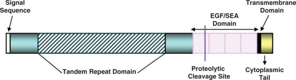
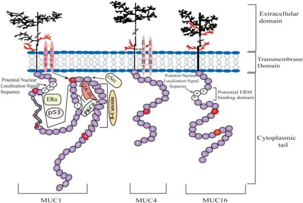
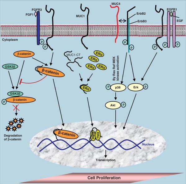
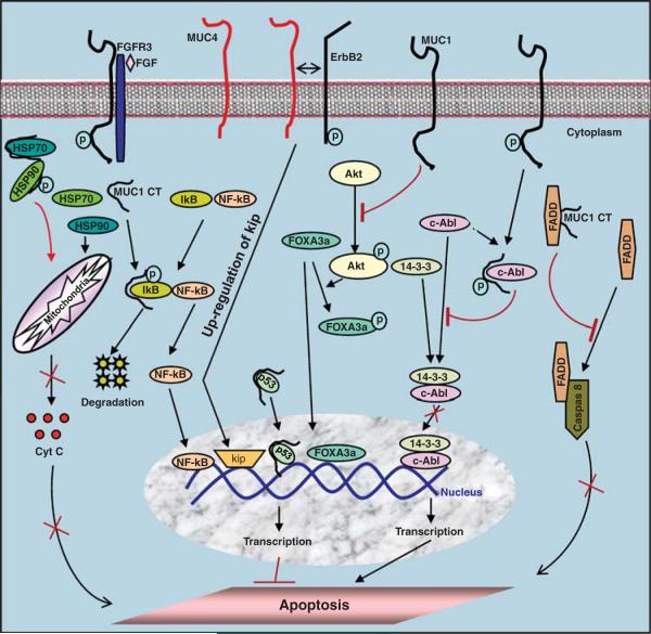
Mucins (MUC) are high molecular weight O-linked glycoproteins whose primary functions are to hydrate, protect, and lubricate the epithelial luminal surfaces of the ducts within the human body. The MUC family is comprised of large secreted gel forming and transmembrane (TM) mucins. MUC1, MUC4, and MUC16 are the well-characterized TM mucins and have been shown to be aberrantly overexpressed in various malignancies including cystic fibrosis, asthma, and cancer. Recent studies have uncovered the unique roles of these mucins in the pathogenesis of cancer. These mucins possess specific domains that can make complex associations with various signaling pathways, impacting cell survival through alterations of cell growth, proliferation, death, and autophagy. The cytoplasmic domain of MUC1 serves as a scaffold for interaction with various signaling proteins. On the other hand, MUC4 mediates its effect by stabilizing and enhancing the activity of growth factor receptor ErbB2. MUC16, previously known as CA125, is a well-known serum marker for the diagnosis of ovarian cancer and has a key role in stimulation and dissemination of ovarian cancer cells by interacting with mesothelin and galectin. Therefore, herein we discuss the function and divergent mechanisms of MUC1, MUC4, and MUC16 in carcinogenesis in the context of alteration in cell growth and survival.


Figures

Figure 1
Figure 1
Prototype structure of the membrane-bound mucins. These mucins typically compose two subunits, based on the putative proteolytic cleavage site. The larger subunit is extracellular and predominantly composed of variable number of tandem repeats. The smaller subunit consists of short extracellular region (containing either sperm protein, enterokinase, and agrin (SEA) domain or epidermal growth factor (EGF)-like domain), single transmembrane domain, and the cytoplasmic tail.
Figure 2
Figure 2
Schematic representation of MUC1, MUC4, and MUC16 cytoplasmic tails (CT). The sequence of cytoplasmic tails of different MUCs is shown with emphasis on tyrosine residue (red) and their known binding partners. MUC1 cytoplasmic tail is the most well-studied and known to interact with proteins with kinase activity (PCKδ, GSK3β, EGFR, and c-Src) and without kinase activity (p53, ERα, β-catenin). The MUC16 cytoplasmic tail has a motif containing polybasic amino-acid sequence, which is a potential site for interaction with cytoskeleton through ERM protein. Both categories of interacting partners are involved in different signaling pathways emphasizing on the critical role of mucin cytoplasmic tail in intracellular signaling events. MUC4 cytoplasmic tail also contains tyrosine residue but till date nothing is known about their intracellular-binding partners. In addition, both MUC1 and MUC16 contain positively charged lysine-arginine rich potential nuclear localization motif in the region juxtaposed to the plasma membrane. In extracellular domain, N and O-linked glycosylations are shown schematically with red and black color, respectively.
Figure 3
Figure 3
Divergent mechanisms of MUC1 and MUC4 for enhanced cancer cell proliferation. MUC1 through its cytoplasmic tail (CT) interacts with the ErbB1 epidermal growth factor receptor tyrosine kinase and increases cell proliferation via activation of extracellular signal-regulated kinases (Erks). MUC4 also contributes to enhanced cellular proliferation through its interaction with another epidermal growth factor receptor tyrosine kinase ErbB2 with subsequent activation of Erk and Akt signaling pathways. By interacting with β-catenin, MUC1-CT inhibits GSK3β-mediated degradation of the β-catenin and increases the expression of cell-cycle progression genes by increasing the nuclear pool of β-catenin. MUC1 also controls cell proliferation by stabilizing estrogen receptor α (ERα).
Figure 4
Figure 4
Illustration of different mechanisms of MUC1 and MUC4 for the repression of apoptosis. MUC1 selectively transactivates p53, FOXA3a, and NF-κB transcription factors and suppresses induction of apoptosis. In contrast, MUC1 blocks nuclear targeting of c-Abl and blocks apoptosis. Activation of MUC1-CT by growth factor signaling causes its binding with HSP90, which transduces signals from the cell membrane to mitochondria that attenuates activation of the intrinsic apoptotic pathway. In addition, MUC1 interacts with the death effector domain of FADD and thereby prevents an activation extrinsic apoptotic pathway. MUC4 has also been proposed to represses apoptosis through the upregulation of kip.

Similar articles

Cited by

References

    1. Agata N, Ahmad R, Kawano T, Raina D, Kharbanda S, Kufe D. MUC1 oncoprotein blocks death receptor-mediated apoptosis by inhibiting recruitment of caspase-8. Cancer Res. 2008;68:6136–6144. - PMC - PubMed
    1. Ahmad R, Raina D, Trivedi V, Ren J, Rajabi H, Kharbanda S, et al. MUC1 oncoprotein activates the IkappaB kinase beta complex and constitutive NF-kappaB signalling. Nat Cell Biol. 2007;9:1419–1427. - PMC - PubMed
    1. Andrianifahanana M, Moniaux N, Schmied BM, Ringel J, Friess H, Hollingsworth MA, et al. Mucin (MUC) gene expression in human pancreatic adenocarcinoma and chronic pancreatitis: a potential role of MUC4 as a tumor marker of diagnostic significance. Clin Cancer Res. 2001;7:4033–4040. - PubMed
    1. Arango ME, Li P, Komatsu M, Montes C, Carraway CA, Carraway KL. Production and localization of Muc4/sialomucin complex and its receptor tyrosine kinase ErbB2 in the rat lacrimal gland. Invest Ophthalmol Vis Sci. 2001;42:2749–2756. - PubMed
    1. Argueso P, Spurr-Michaud S, Russo CL, Tisdale A, Gipson IK. MUC16 mucin is expressed by the human ocular surface epithelia and carries the H185 carbohydrate epitope. Invest Ophthalmol Vis Sci. 2003;44:2487–2495. - PubMed

Publication types