. 2010 May 27;29(21):3044-53.
doi: 10.1038/onc.2010.78. Epub 2010 Mar 29.

SSBP2 is an in vivo tumor suppressor and regulator of LDB1 stability

Affiliations

SSBP2 is an in vivo tumor suppressor and regulator of LDB1 stability

Y Wang et al. Oncogene. .

Abstract

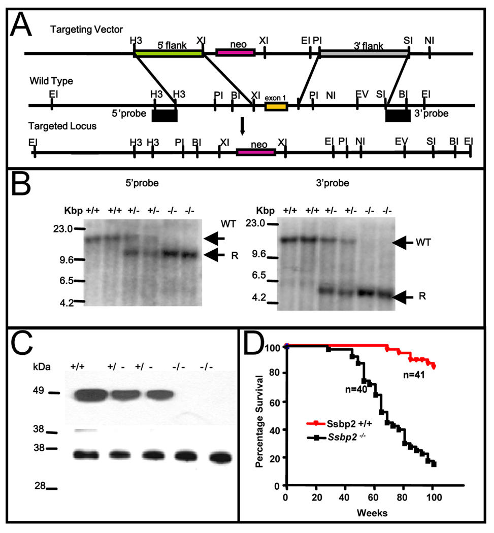
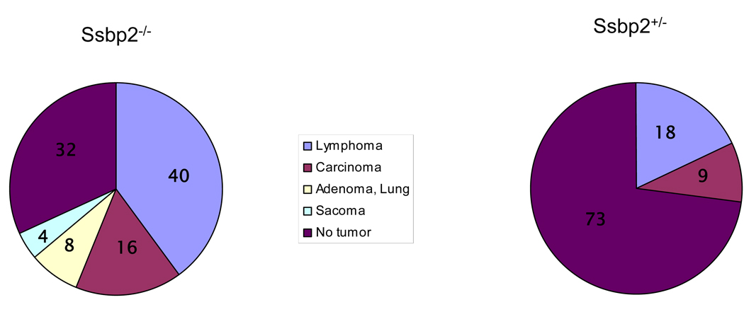
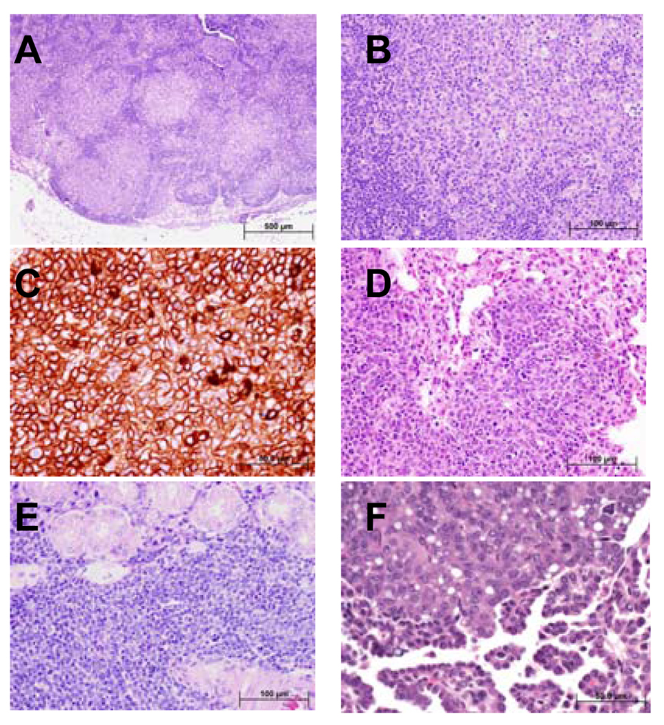
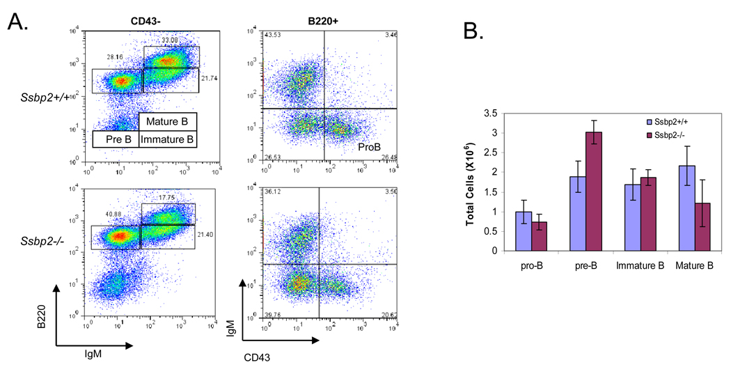
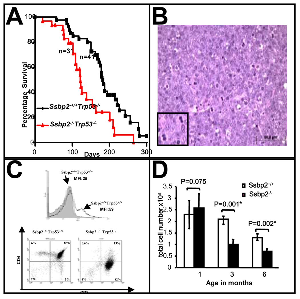
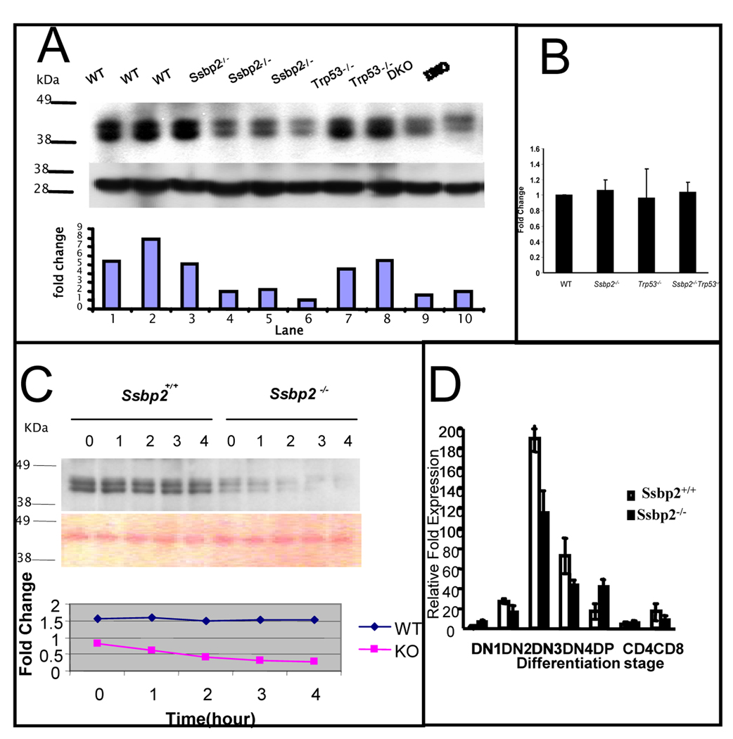
SSBP proteins bind and stabilize transcriptional cofactor LIM domain-binding protein1 (LDB1) from proteosomal degradation to promote tissue-specific transcription through an evolutionarily conserved pathway. The human SSBP2 gene was isolated as a candidate tumor suppressor from a critical region of loss in chromosome 5q14.1. By gene targeting, we show increased predisposition to B-cell lymphomas and carcinomas in Ssbp2(-/-) mice. Remarkably, loss of Ssbp2 causes increased LDB1 turnover in the thymus, a pathway exploited in Trp53(-/-)Ssbp2(-/-) mice to develop highly aggressive, immature thymic lymphomas. Using T-cell differentiation as a model, we report a stage-specific upregulation of Ssbp2 expression, which in turn regulates LDB1 turnover under physiological conditions. Furthermore, transcript levels of pTalpha, a target of LDB1-containing complex, and a critical regulator T-cell differentiation are reduced in Ssbp2(-/-) immature thymocytes. Our findings suggest that disruption of the SSBP2-regulated pathways may be an infrequent but critical step in malignant transformation of multiple tissues.


Conflict of interest statement

Conflict of Interest

The authors do not have any competing financial interest in the results presented.

Figures

Figure 1
Figure 1. Targeted disruption of the murine Ssbp2 locus
(A) Partial restriction maps of the targeting vector, mouse Ssbp2 genomic locus and the expected targeted allele are depicted. Ssbp2 exon 1 containing the initiation ATG was replaced with a neomycin resistance gene in the plasmid vector pPNT1. Unique single copy fragments flanking the integration are shown. The restriction sites indicated are: BI (BamH1), EI (Eco RI), EV (Eco RV), H3 (HindIII) NI (NotI), PI (PstI), SI (SpeI) and XI (XhoI). The probes used were derived from the 5’ HindIII-HindIII and 3’ SpeI-BamHI fragments. These probes detected different sized wild type and rearranged fragments containing the inserted neo casette in DNAs digested with Eco RI (B) Southern blot analysis with 5’ probe or 3’ probe of Eco RI-digested tail DNA from littermates obtained from intercrossing Ssbp2+/− mice. Germline DNA yielded a 13.9 kbp fragment when cleaved with EcoRI. The knockout allele gave 10.1 kbp and 4.5 kbp fragments, respectively, with the 5’ and 3’ probes. Unique wild type (WT) and recombinant (R) fragments are denoted. (C) Immunoblotting of nuclear lysate from Ssbp2+/+, Ssbp2+/− and Ssbp2−/− mice. 30 µg of protein aliquots from nuclear lysates of 4 week-old mice thymi were separated on a 4–12% NuPage gel, transferred to a Hybond-P PVDF membrane, and probed with SSBP2 antibody. Lower panel shows Ponceau staining carried out to visualize overall protein loading. (D) Kaplan-Meier survival curve showing the percent survival of Ssbp2 −/− (n=41) and Ssbp2 +/+ (n=40) cohort mice as a function of age in weeks. Both groups of mice were monitored for greater than two years and sacrificed when moribund. Survival of SSBP2+/+ and SSBP2−/− differed significantly (P < 0.0001).
Figure 2
Figure 2. Tumor spectrum in Ssbp2 null mice
A) Pie chart depicts relative incidence of malignancies seen in Ssbp2−/− mice. A total of 14 tumors from 25 mice ( Supplementary Table II) was used to determine the frequencies. B) Eleven age matched Ssbp2+/− mice were sacrificed for total necropsy. Three malignancies were found in two mice. Numbers inside each slice denote the percentage of specific tumor type.
Figure 3
Figure 3. Ssbp2−/− mice develop disseminated high-grade large B-cell lymphoma and other malignancies
A total of 25 moribund, age matched non-morbid mice were necropsied (Table II). (A) Histological sections from mandibular lymph node demonstrate total effacement of the normal architecture by neoplastic lymphoid follicles that lack the normal structure of the reactive lymphoid follicles including distinctive germinal centers. The neoplastic lymphoid follicles are arranged in a back-to-back pattern without significant interfollicular lymphoid tissue. (B) At higher magnification the cells within the neoplastic follicles are intermediate to large in size with round vesicular nuclei, occasional prominent 1–3 nucleoli, and abundant eosinophilic cytoplasm. Patchy increased mitotic figures are present. C. Immunohistochemical staining where the neoplastic lymphocytes are strongly positive for B220 supporting a B-cell immunophenotype. The histological and cytological features are most consistent with high-grade large B-cell lymphoma of follicle center cell origin. D Sections from the lung showing that most of the alveolar spaces are lacking because of extensive infiltration of the alveolar walls by the high-grade large B-cell lymphoma cells. The cytological features of the lymphoma cells are similar to the ones described in the lymph node shown in B. E. Extensive interstitial and diffuse infiltration of salivary gland by malignant lymphoma cells. The neoplastic lymphoma cells focally invade the glandular epithelial structures. The cytological features of the lymphoma cells are similar to the ones described in the lymph node shown in (B). F. Ssbp2−/− mice also developed carcinomas such as this low-grade adenocarcinoma of lung. The tumor is composed of bland glandular structures lined with cuboidal to low columnar cells that invade the pulmonary stroma. Bars denote the magnification.
Figure 4
Figure 4. Expansion of the preB cell compartment in Ssbp2−/− bone marrow
A. FACS Analysis of Pro-B cells (B220+CD43+IgM), preB cells (B220+CD43IgM), immature B cells (B220low CD43 IgM+), and mature B cells (B220 hi CD43 IgM+ ) in bone marrow harvests cells from wild type and Ssbp2−/− mice. Data shown are representative flow cytometric analyses of five independent experiments. B. Summary of results shown in Fig. 4A.
Figure 5
Figure 5. Loss of Ssbp2 exacerbates aggressive thymic lymphomas in Trp53−/− mice
(A) Kaplan- Meir survival plot of Ssbp2 −/− Trp53−/−, Ssbp2 +/+ Trp53−/− mice. Cohorts of Ssbp2 −/− Trp53−/− (n=31), Ssbp2 +/+ Trp53−/− (n=41) were monitored daily for 300 days for symptoms of disease and moribund mice were euthanized. The median survival was 122 days for the Ssbp2 −/− Trp53−/− mice compared to 182 days for the Ssbp2+/+ Trp53−/− mice. Statistical analysis (by Prism3 software) confirmed the difference in survival between the two groups to be significant (p=0.0019). (B) Histological characteristics of thymic lymphomas in Ssbp2 −/− Trp53−/− mice Hematoxylin-Eosin stain of thymic tumor from a double knockout mouse. The lymphoma is composed of large lymphoid cells with high mitotic index. Inset shows representative mitotic nuclei. The neoplastic lymphocytes are round with vesicular nuclei, single centrally located nucleoli, and scant eosinophilic cytoplasm. Bar denotes the scale. (C) Ssbp2 −/− Trp53 −/− thymic lymphomas are CD3lo ,CD4lowCD8+ (A) The gray shading shows expression of CD3 in the Ssbp2 −/−, Trp53−/− thymic lymphoma cells, compared with that of the Ssbp2 +/+, Trp53+/+ mouse. (B) The right panel is a representative for Ssbp2 −/−, Trp53−/− thymic lymphoma, in which most of the cells are CD8+, CD4 low. The left panel is a wild type control (Ssbp2 +/+, Trp53+/+) thymus in which most of the cells are CD4+,CD8+. (D) Thymic hypoplasia is accelerated in Ssbp2−/− mice Thymi from Ssbp2+/+ and Ssbp2−/− mice were harvested at the indicated time points, minced, single cells isolated and counted. Each group includes a minimum of four mice.
Figure 6
Figure 6. Decreased LDB1 half life in Ssbp2−/− thymocytes underlies impaired T cell differentiation
(A) LDB1 levels are decreased in the thymi of Ssbp2−/− mice. Nuclear proteins from 4 weeks old wild type, Ssbp2 −/−, Trp53−/− and Ssbp2 −/− Trp53−/− mice thymi were separated on a 4–12% NuPage gel, transferred to nitrocellulose and probed with Ldb1 antibody. Lower panel shows Ponceau staining to denote equal loading. The signals were analyzed by IMAGE analysis software and the ratio for each sample is denoted. Representative samples from triplicate experiments are shown. (B) Ldb1 transcript levels are not altered in Ssbp2−/− thymocytes Ssbp2 transcripts in four weeks old thymi were estimated by Real-time PCR. Results show that Ldb1 transcript levels were similar between all the four genotypes tested. Data represent average of two separate experiments each with triplicate reactions. (C) LDB1 half life is shortened in the absence of Ssbp2 Short term cultured thymocytes from wild type and Ssbp2−/− mice were treated with cycloheximide for the indicated lengths of time and whole cell lysates isolated were examined for LDB1 levels by immunoblotting. Protein loading was quantified by Ponceau staining. The signals were analyzed by IMAGE analysis software and the ratios at various time points plotted to determine LDB1 half life. (D) preTα expression is decreased in Ssbp2−/− thymus. Thymocytes from four weeks old mice were divided into seven subsets based on maturity. Real-time PCRs were performed on purified cell populations using specific primers for pTα. For each cDNA pool, transcript levels were normalized against 18sRNA within that sample. Two separate experiments, each with triplicate reactions were done.

References

    1. Agulnick AD, Taira M, Breen JJ, Tanaka T, Dawid IB, Westphal H. Nature. 1996;384:270–272. - PubMed
    1. Aoyama M, Ozaki T, Inuzuka H, Tomotsune D, Hirato J, Okamoto Y, Tokita H, Ohira M, Nakagawara A. Cancer Res. 2005;65:4587–4597. - PubMed
    1. Bach I, Rodriguez-Esteban C, Carriere C, Bhushan A, Krones A, Rose DW, Glass CK, Andersen B, Izpisua Belmonte JC, Rosenfeld MG. Nat Genet. 1999;22:394–399. - PubMed
    1. Baseta JG, Stutman O. J Immunol. 2000;165:5621–5630. - PubMed
    1. Cai Y, Xu Z, Nagarajan L, Brandt SJ. Biochem Biophys Res Commun. 2008;373:303–308. - PMC - PubMed

Publication types