Review
. 2010 Jul;48(8):2303-15.
doi: 10.1016/j.neuropsychologia.2010.03.018. Epub 2010 Mar 27.

Spatial working memory deficits in GluA1 AMPA receptor subunit knockout mice reflect impaired short-term habituation: evidence for Wagner's dual-process memory model

Affiliations
Review

Spatial working memory deficits in GluA1 AMPA receptor subunit knockout mice reflect impaired short-term habituation: evidence for Wagner's dual-process memory model

David J Sanderson et al. Neuropsychologia. 2010 Jul.

Abstract

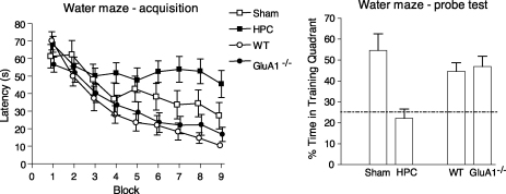
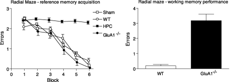
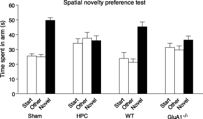
Genetically modified mice, lacking the GluA1 AMPA receptor subunit, are impaired on spatial working memory tasks, but display normal acquisition of spatial reference memory tasks. One explanation for this dissociation is that working memory, win-shift performance engages a GluA1-dependent, non-associative, short-term memory process through which animals choose relatively novel arms in preference to relatively familiar options. In contrast, spatial reference memory, as exemplified by the Morris water maze task, reflects a GluA1-independent, associative, long-term memory mechanism. These results can be accommodated by Wagner's dual-process model of memory in which short and long-term memory mechanisms exist in parallel and, under certain circumstances, compete with each other. According to our analysis, GluA1(-/-) mice lack short-term memory for recently experienced spatial stimuli. One consequence of this impairment is that these stimuli should remain surprising and thus be better able to form long-term associative representations. Consistent with this hypothesis, we have recently shown that long-term spatial memory for recently visited locations is enhanced in GluA1(-/-) mice, despite impairments in hippocampal synaptic plasticity. Taken together, these results support a role for GluA1-containing AMPA receptors in short-term habituation, and in modulating the intensity or perceived salience of stimuli.


Figures

Fig. 1
Fig. 1
Hippocampal lesions, but not GluA1 deletion impair performance of spatial reference memory in the water maze task. Water maze–acquisition: mean escape latency (±S.E.M.) to find the platform across nine days of training for sham (Sham) and hippocampal lesioned mice (HPC), and for wild-type (WT) and GluA1−/− mice. Water maze–probe test: mean percent time in the training quadrant (±S.E.M.) during a probe trial in which the platform is removed from the pool and the mice are allowed to swim freely. The dashed line indicates chance performance. Sham and hippocampal lesioned mice results reproduced from Deacon et al. (2002). Wild-type and GluA1−/− mice results reproduced from Reisel et al. (2002).
Fig. 2
Fig. 2
Hippocampal lesions, but not GluA1 knockout impair acquisition of the spatial reference memory component of the radial maze task. Radial-arm maze–reference memory acquisition: mean number of reference memory errors per trial (±S.E.M.) during 6 blocks of training (4 trials per block). Mice were trained to discriminate between 3 baited arms and 3 non-baited arms of a 6 arm radial maze. Doors prevented mice re-entering arms they had already visited on that particular visit to the maze (i.e. prevented working memory errors) during this stage of the experiment. Sham lesioned (Sham), wild-type (WT), and Glua1−/− mice all acquired the task at a similar rate. Hippocampal lesioned mice (HPC) were completely unable to learn which arms were baited and which arms were not baited. Radial-arm maze–working memory performance: mean number of working memory errors per trial (±S.E.M.) for wild-type (WT) and Glua1−/− mice. During this stage of testing mice were still rewarded in the same 3 arms of the maze and not rewarded in the 3 non-baited arms, but now they were allowed to re-enter arms as often as they liked, and rewards were not replaced within a trial. Data reproduced from Schmitt et al. (2003).
Fig. 3
Fig. 3
Hippocampal lesions and GluA1 knockout both impair performance on a spatial novelty preference test. Mean time spent in arms (±S.E.M.). Sham and wild-type (WT) mice exhibit a preference for a previously unexposed (Novel) arm of a Y-maze over two familiar arms to which they have previously been exposed (Start and Sample). GluA1 knockout mice (GluA1−/−) and hippocampal lesioned mice (HPC) did not show a significant preference for the novel arm. Data reproduced from Sanderson et al. (2007).
Fig. 4
Fig. 4
The states of activation, which elements of a memory can reside, and the permissible transitional routes between states, according to Wagner (1981). Presentation of a stimulus leads to a proportion of its elements transferring from the inactive state (I) to the A1 state (route 1). Elements then decay to a secondary activation state, A2 (route 2), before returning to an inactive state, I (route 3). Elements that are active in the A2 state cannot return to the A1 state on subsequent presentation of the stimulus, thus leading to reduced A1 activity. A2 state activity can occur due to the recent presentation of a stimulus (self-generated priming; route 2). Also, presentation of a stimulus leads to A2 state activation of elements of other stimuli with which it is associated (retrieval-generated priming; route 4).
Fig. 5
Fig. 5
The design of Experiments 1 and 2 in Sanderson et al. (2009). (a) During exposure training mice were allowed to explore the Start arm and the Sample arm for five 2-min trials. Access to the Novel arm was blocked. During the novelty preference test mice were allowed to explore the two familiar arms (Start and Sample) and the previously unvisited, Novel arm for a period of 2 min. (b) In Experiment 1 the interval between exposure trials (represented by the black bars) and also the interval prior to the novelty preference test (represented by the white bars) was either 1 min (1 min ITI) or 24 h (24 h ITI). (c) In Experiment 2, two groups of mice from each genotype received exposure training with a 1 min interval between trials and two further groups from each genotype received exposure training with a 24 h interval between trials. One group from each training condition received the novelty preference 1 min after the last training trial. The other group received the test 24 h after the last training trial.
Fig. 6
Fig. 6
GluA1 knockout impairs short-term spatial novelty preference, but enhances long-term novelty preference. The preference for the Novel arm over the Familiar (Sample) arm is shown as a discrimination ratio (Novel/[Novel + Familiar]). Scores greater than 0.5 indicate a novelty preference. The dashed lines indicate chance performance. Errors bars indicate ±S.E.M. (a) In Experiment 1 (see Fig. 5b) GluA1−/− mice (KO) were enhanced in the long, 24 h ITI condition relative to wild-type mice (WT), but impaired in the short, 1 min ITI condition. (b) Hippocampal lesioned mice (Hpc) failed to show a significant novelty preference in either condition. (c) In Experiment 2 (see Fig. 5c) GluA1−/− mice were enhanced when the training trials were separated by 24 h. There was no significant interaction between genotype and the test interval. (d) The results of Experiment 2 collapsed across the short, 1 min and long, 24 h test intervals to show the independent effects of the training ITI in wild-type and GluA1−/− mice. Data reproduced from Sanderson et al. (2009).
Fig. 7
Fig. 7
Simulations of Wagner's SOP model (1981). To simulate the effect of GluA1 deletion on spatial novelty preference, calculations were performed to determine the difference in the memory states following two exposure training trials followed by a novelty test. It is assumed that during an exposure trial there are elements of a stimulus that are active throughout the trial (e.g. a CS) that can form an association, and thus predict the occurrence of elements of another stimulus that are only active towards the end of the trial (e.g. a US). This may, for example, describe the possible associative learning between spatial stimuli experienced in the Start arm and spatial stimuli experienced in the Sample arm. The CS is a 10-moment stimulus and the US is a 5 moment stimulus that co-terminated. The short and long training ITI were a length of 5 and 100 moments respectively. The novelty preference test consisted of 6 moments of the CS and 1 moment of the trained US and a novel US that co-terminated. For all stimuli the intensity parameter was 0.2. For normal wild-type mice the decay parameters for A1 (pd1) and A2 (pd2) were 0.2 and 0.04 respectively. For GluA1−/− mice the value of pd1 was reduced to 0.11. The excitatory learning rate parameter was 0.07 and the inhibitory learning rate parameter was 0.014 (for other details see Sanderson et al., 2009). Simulations were carried out for training with a short and a long ITI and with testing after a short or long interval for both conditions. The results are subsequently collapsed across testing conditions so as to show the independent effects of the training ITI. (a) The difference in A1 activity between a novel US and a trained US. A positive value indicates greater relative novel US A1 activity. When pd1 = 0.2, a short training ITI causes a greater relative novel US A1 activity than a long training ITI. However, when pd1 = 0.11, a long training ITI causes a greater relative novel US A1 activity than a short training ITI. (b) The momentary change in net associative strength (ΔV) over two exposure training trials. A slower A1 to A2 decay rate (pd1 = 0.11) causes greater increments in associative strength than a faster decay rate (pd1 = 0.2) by increasing the concurrent CS and US A1 state activity.

Similar articles

Cited by

References

    1. Aggleton J.P., Neave N., Nagle S., Sahgal A. A comparison of the effects of medial prefrontal, cingulate cortex, and cingulum bundle lesions on tests of spatial memory: Evidence of a double dissociation between frontal and cingulum bundle contributions. Journal of Neuroscience. 1995;15(11):7270–7281. - PMC - PubMed
    1. Aggleton J.P., Pearce J.M. Neural systems underlying episodic memory: Insights from animal research. Philosophical Transactions of the Royal Society of London. Series B: Biological Sciences. 2001;356(1413):1467–1482. - PMC - PubMed
    1. Andrasfalvy B.K., Smith M.A., Borchardt T., Sprengel R., Magee J.C. Impaired regulation of synaptic strength in hippocampal neurons from GluR1-deficient mice. Journal of Physiology. 2003;552(Pt 1):35–45. - PMC - PubMed
    1. Baddeley A.D., Hitch G. Working memory. In: Bower G.H., editor. Vol. 8. Academic Press; New York: 1974. pp. 47–89. (The psychology of learning and motivation: Advances in research and theory).
    1. Bannerman D.M., Deacon R.M., Brady S., Bruce A., Sprengel R., Seeburg P.H. A comparison of GluR-A-deficient and wild-type mice on a test battery assessing sensorimotor, affective, and cognitive behaviors. Behavioral Neuroscience. 2004;118(3):643–647. - PubMed

Substances