. 2010 May 15;588(Pt 10):1683-93.
doi: 10.1113/jphysiol.2009.183418. Epub 2010 Mar 29.

Direct voltage control of endogenous lysophosphatidic acid G-protein-coupled receptors in Xenopus oocytes

Affiliations

Direct voltage control of endogenous lysophosphatidic acid G-protein-coupled receptors in Xenopus oocytes

Juan Martinez-Pinna et al. J Physiol. .

Abstract

Lysophosphatidic acid (LPA) G-protein-coupled receptors (GPCRs) play important roles in a variety of physiological and pathophysiological processes, including cell proliferation, angiogenesis, central nervous system development and carcinogenesis. Whilst many ion channels and transporters are recognized to be controlled by a change in cell membrane potential, little is known about the voltage dependence of other proteins involved in cell signalling. Here, we show that the InsP(3)-mediated Ca(2+) response stimulated by the endogenous LPA GPCR in Xenopus oocytes is potentiated by membrane depolarization. Depolarization was able to repetitively stimulate transient [Ca(2+)](i) increases after the initial agonist-evoked response. In addition, the initial rate and amplitude of the LPA-dependent Ca(2+) response were significantly modulated by the steady holding potential over the physiological range, such that the response to LPA was potentiated at depolarized potentials and inhibited at hyperpolarized potentials. Enhancement of LPA receptor-evoked Ca(2+) mobilization by membrane depolarization was observed over a wide range of agonist concentrations. Importantly, the amplitude of the depolarization-evoked intracellular Ca(2+) increase displayed an inverse relationship with agonist concentration such that the greatest effect of voltage was observed at near-threshold levels of agonist. Voltage-dependent Ca(2+) release was not induced by direct elevation of InsP(3) or by activation of heterotrimeric G-proteins in the absence of agonist, indicating that the LPA GPCR itself represents the primary site of action of membrane voltage. This novel modulation of LPA signalling by membrane potential may have important consequences for control of Ca(2+) signals both in excitable and non-excitable tissues.


Figures

Figure 4
Figure 4. Functional InsP3 receptors and phospholipase-C activation are required for the voltage control of Ca2+ release during LPA receptor stimulation
A, effect of depolarization (−80 to −10 mV) on [Ca2+]i in the presence of 10 nm LPA alone (first voltage step) or following co-application of 100 μm 2-APB, an inhibitor of InsP3 receptors (second voltage step). B, amplitude of the depolarization-evoked Ca2+ response (−80 to −10 mV, 60 s) in the presence of LPA after incubation with the phospholipase C inhibitor U-73122 (10 μm, filled circles), for different durations or following exposure to either its inactive analogue U-73343 (10 μm, open circles) or to DMSO at the same concentration used to dissolve these drugs (1/100 dilution, open triangles), for the duration required to observe the maximal effect with U-73122 (60–65 min). All experiments were performed in Ca2+-free external solution. Each point represents a different cell.
Figure 1
Figure 1. Effect of membrane voltage on LPA-evoked Ca2+ responses in Xenopus oocytes
A–E, intracellular Ca2+ recordings from oocytes under two-electrode voltage clamp in normal (Ca2+-containing) Ringer solution (A–C) or Ca2+-free Ringer solution (D and E). Upper panels show the Fluo5, f/f0 ratio and lower panels the membrane voltage. A, typical recording showing the lack of effect of a 70 mV depolarizing voltage step (−80 to −10 mV, lower panel) on [Ca2+]i (top panel) in the absence of agonist and the typical response to 10 nm LPA held at −80 mV. B and C, typical recordings of the [Ca2+]i increase (B, 75% of cells) and [Ca2+]i decrease (C, 25% of cells) evoked by depolarization during exposure to LPA. D, recording showing the repetitive and reversible nature of the depolarization-evoked Ca2+ increase observed in 100% of oocytes in Ca2+-free medium. E, effect of membrane hyperpolarization (−50 to −120 mV) on the response to 10 nm LPA. F, bar graph showing the average depolarization-induced Ca2+ response (Δf/f0) in the absence (no agonist) or presence of 10 nm LPA in the presence and absence of external Ca2+ and in the presence of 5 mm Ni2+ in Ca2+-free Ringer solution. The depolarization-evoked response in Ca2+-free Ringer solution was significantly smaller than that observed in normal Ringer solution (asterisk, P < 0.05).
Figure 2
Figure 2. Effect of the holding potential on LPA-evoked Ca2+ responses
A and B, representative traces showing the influence of the holding potential on LPA-evoked Ca2+ release at −80 and −20 mV, respectively, in a Ca2+-free external solution. Time scale in A is the same as in B. The lines with arrows in A indicate the parameters quantified in panels C, D and E. The latency (C), the time to peak (D) and the amplitude (E) of LPA-evoked Ca2+ response were measured in 8–9 oocytes at the constant holding potential shown (−80, −50 and −20 mV). The values for the three parameters were significantly different (asterisk, P < 0.05) between holding potentials of −80 and −20 mV.
Figure 3
Figure 3. Potentiation of LPA receptor-evoked Ca2+ response by depolarization is greatest at low concentrations of LPA
AC, effect of a depolarizing pulse (−80 to −10 mV) on [Ca2+]i at a subthreshold (0.1 nm, A), near-threshold (1 nm, B) and suprathreshold (100 nm, C) concentration of LPA in Ca2+-free external solution. At 0.1 nm LPA (A), all oocytes failed to respond to the agonist alone, whereas 3 out of 8 cells displayed a subsequent depolarization-evoked [Ca2+]i increase. D, semilogarithmic concentration–response relationship for the depolarization-evoked [Ca2+]i increase (open circles, measured as a percentage of the corresponding LPA-evoked response) and LPA-evoked peak response (Δf/f0, filled squares), versus the LPA concentration. Potentiation by depolarization at 0.1 nm LPA is excluded due to lack of agonist response. LPA-evoked peak response data were best fitted with the logistic equation: formula image
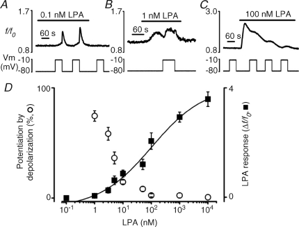
, where x0 is the middle value, p is the power and A1 and A2 are the initial and final Y values, respectively. Each point is the mean and s.e.m. of 4–13 cells.
Figure 5
Figure 5. Lack of depolarization-evoked Ca2+ response following direct elevation of InsP3 or the heterotrimeric G-protein activation with aluminium tetrafluoride
A, lack of effect of depolarizing pulses (lower panel) on [Ca2+]i (top panel) during the response to ∼500 fmol of intracellularly injected InsP3 (see Methods). Arrow indicates the point of InsP3 injection. B, cells were exposed to the heterotrimeric G-protein activator AlF4 using a mixture of 10 mm NaF and 200 μm AlCl3. Exposure to AlF4 reversibly induced a [Ca2+]i increase (top panel) but depolarizing pulses (lower panel) had no effect on [Ca2+]i. All experiments were performed in Ca2+-free external solution.

References

    1. An S, Bleu T, Zheng Y, Goetzl EJ. Recombinant human G protein-coupled lysophosphatidic acid receptors mediate intracellular calcium mobilization. Mol Pharmacol. 1998;54:881–888. - PubMed
    1. Baker DL, Desiderio DM, Miller DD, Tolley B, Tigyi GJ. Direct quantitative analysis of lysophosphatidic acid molecular species by stable isotope dilution electrospray ionization liquid chromatography-mass spectrometry. Anal Biochem. 2001;292:287–295. - PubMed
    1. Ben Chaim Y, Chanda B, Dascal N, Bezanilla F, Parnas I, Parnas H. Movement of ‘gating charge’ is coupled to ligand binding in a G-protein-coupled receptor. Nature. 2006;444:106–109. - PubMed
    1. Ben Chaim Y, Tour O, Dascal N, Parnas I, Parnas H. The M2 muscarinic G-protein-coupled receptor is voltage-sensitive. J Biol Chem. 2003;278:22482–22491. - PubMed
    1. Berridge MJ. Inositol trisphosphate and calcium signalling. Nature. 1993;361:315–325. - PubMed

Publication types

LinkOut - more resources