

doi: 10.1074/jbc.R110.117671.
Epub 2010 Mar 29.
Mechanism of intraparticle synthesis of the rotavirus double-stranded RNA genome
Affiliations
- PMID: 20351108
- PMCID: PMC2881735
- DOI: 10.1074/jbc.R110.117671
Item in Clipboard
Mechanism of intraparticle synthesis of the rotavirus double-stranded RNA genome
J Biol Chem.
.
Abstract
Rotaviruses perform the remarkable tasks of transcribing and replicating 11 distinct double-stranded RNA genome segments within the confines of a subviral particle. Multiple viral polymerases are tethered to the interior of a particle, each dedicated to a solitary genome segment but acting in synchrony to synthesize RNA. Although the rotavirus polymerase specifically recognizes RNA templates in the absence of other proteins, its enzymatic activity is contingent upon interaction with the viral capsid. This intraparticle strategy of RNA synthesis helps orchestrate the concerted packaging and replication of the viral genome. Here, we review our current understanding of rotavirus RNA synthetic mechanisms.
Figures
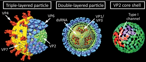
Structural organization of the RV virion as determined by cryoelectron microscopy and image reconstruction. Left, cutaway of the TLP, identifying the outer capsid attachment protein VP4, glycoprotein VP7, and the internal VP6 and VP2 protein layers. Middle, the DLP, which has been colored based on radial distance from the center of the particle. Part of the VP2 and VP6 layers has been computationally removed to reveal the tubular organization of dsRNA spooled around internal projections formed at 5-fold vertices by VP2 N termini, VP1, and VP3. Right, the VP2 core shell, which is shown primarily in green, with VP2-A monomers encircling one 5-fold vertex shown in red and VP2-B monomers encircling one 3-fold vertex shown in purple. A single asymmetrical VP2 dimer formed by the interaction of a VP2-A monomer of one vertex and the VP2-B monomer of another vertex is outlined in white. The location of a type I channel is indicated. Images were kindly provided by B. V. V. Prasad (Baylor College of Medicine).
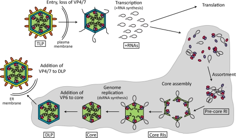
Model of RNA synthesis during the RV life cycle. During entry into target cells, VP4 (orange) and VP7 (yellow) are lost from TLPs. In the resultant DLPs, PCs composed of VP1 (purple) and VP3 (pink) catalyze synthesis of multiple copies of each of the 11 species of RV +RNAs (black ellipses), which anneal in cis to form panhandle structures, with the 3′CS+ remaining largely unbase-paired. The +RNAs serve as templates for translation or are packaged into assembling particles. As viral proteins and +RNAs accumulate in the viroplasm (gray), VP1, VP3, and +RNAs associate to form precore RIs, followed by addition of VP2 (green) to form catalytically active core RIs. Genome replication results in synthesis of the 11 genomic dsRNA segments. VP6 (blue) is added to mature cores, forming DLPs. Finally, DLPs move into the endoplasmic reticulum (ER) where, by budding, they acquire the outer VP4 and VP7 layer.
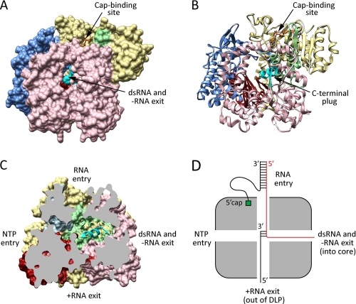
Structural organization of the RV RdRp. A and B, surface and ribbon representations of VP1 (Protein Data Bank code 2R7R). A m7G cap (orange sticks) is shown in the putative cap-binding site. C, cutaway of VP1 (rotated 90° to the right relative to the image in A) showing the four-tunnel architecture of the polymerase. Putative functions of the tunnels are indicated. For A–C, the N-terminal domain of VP1 is colored yellow; the C-terminal domain is colored pink; and the fingers, palm, and thumb subdomains of the polymerase domain are colored blue, red, and green, respectively. The C-terminal plug is colored cyan. D, schematic depicting the four tunnels of VP1, oriented as in C, during transcription. +RNA is colored black, and −RNA is colored red. The 5′-cap (green) of the plus strand of a dsRNA template undergoing transcription is shown anchored into the cap-binding site.
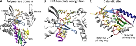
VP1 RNA recognition and polymerization. Shown are ribbon drawings of the polymerase domain (A), template recognition region (B), and catalytic site (C) of VP1 (Protein Data Bank code 2R7R). In all images, a bound oligonucleotide (3′CS+, 5′-UGUGACC-3′; yellow sticks) is shown, and RdRp motifs are colored as follows: motif A (pink), motif B (purple), motif C (green), motif D (navy), motif E (yellow), and motif F (light blue). The remaining portions of VP1 are colored gray. In B, nucleotide bases of the 3′CS+ are labeled, and side chains of VP1 residues that interact with the RNA are shown. Hydrogen bonds formed with the RNA bases and ribose phosphate backbone are shown with green and red lines, respectively. In A and C, the RV priming loop is colored red. In C, nucleotide bases of the 3′-terminal ACC stack are labeled, and the catalytic aspartates of motifs A and C are shown. NTPs (green sticks), divalent cations (slate-blue spheres), and the priming loop (gold) from the structure of reovirus λ3 RdRp (Protein Data Bank code 1N1H) have been overlaid to highlight the overshot position of the RNA and the differences in priming loop orientation of the two polymerases.
Similar articles
-
Assortment and packaging of the segmented rotavirus genome.Trends Microbiol. 2011 Mar;19(3):136-44. doi: 10.1016/j.tim.2010.12.002. Epub 2010 Dec 31. Trends Microbiol. 2011. PMID: 21195621 Free PMC article. Review.
-
In Vitro Double-Stranded RNA Synthesis by Rotavirus Polymerase Mutants with Lesions at Core Shell Contact Sites.J Virol. 2019 Sep 30;93(20):e01049-19. doi: 10.1128/JVI.01049-19. Print 2019 Oct 15. J Virol. 2019. PMID: 31341048 Free PMC article.
-
Rotavirus RNA polymerase requires the core shell protein to synthesize the double-stranded RNA genome.J Virol. 1997 Dec;71(12):9618-26. doi: 10.1128/JVI.71.12.9618-9626.1997. J Virol. 1997. PMID: 9371626 Free PMC article.
-
Rotavirus replication: plus-sense templates for double-stranded RNA synthesis are made in viroplasms.J Virol. 2004 Jul;78(14):7763-74. doi: 10.1128/JVI.78.14.7763-7774.2004. J Virol. 2004. PMID: 15220450 Free PMC article.
-
Rotavirus NSP2: A Master Orchestrator of Early Viral Particle Assembly.Viruses. 2024 May 21;16(6):814. doi: 10.3390/v16060814. Viruses. 2024. PMID: 38932107 Free PMC article. Review.
Cited by
-
Mycoreovirus genome alterations: similarities to and differences from rearrangements reported for other reoviruses.Front Microbiol. 2012 Jun 1;3:186. doi: 10.3389/fmicb.2012.00186. eCollection 2012. Front Microbiol. 2012. PMID: 22675320 Free PMC article.
-
Interaction between a Unique Minor Protein and a Major Capsid Protein of Bluetongue Virus Controls Virus Infectivity.J Virol. 2018 Jan 17;92(3):e01784-17. doi: 10.1128/JVI.01784-17. Print 2018 Feb 1. J Virol. 2018. PMID: 29142128 Free PMC article.
-
Structure of the T=13 capsid of infectious pancreatic necrosis virus (IPNV)-a salmonid birnavirus.J Virol. 2025 Feb 25;99(2):e0145424. doi: 10.1128/jvi.01454-24. Epub 2025 Jan 16. J Virol. 2025. PMID: 39817769 Free PMC article.
-
The battle between rotavirus and its host for control of the interferon signaling pathway.PLoS Pathog. 2013 Jan;9(1):e1003064. doi: 10.1371/journal.ppat.1003064. Epub 2013 Jan 24. PLoS Pathog. 2013. PMID: 23359266 Free PMC article. Review.
-
High-resolution comparative atomic structures of two Giardiavirus prototypes infecting G. duodenalis parasite.PLoS Pathog. 2024 Apr 10;20(4):e1012140. doi: 10.1371/journal.ppat.1012140. eCollection 2024 Apr. PLoS Pathog. 2024. PMID: 38598600 Free PMC article.
References
-
- Mertens P. P., Diprose J. (2004) Virus Res. 101, 29–43 - PubMed
-
- Patton J. T., Vasquez-Del Carpio R., Tortorici M. A., Taraporewala Z. F. (2007) Adv. Virus Res. 69, 167–201 - PubMed
-
- McDonald S. M., Patton J. T. (2009) in Viral Genome Replication ( Cameron C. E., Gotte M., Raney K. D. eds) pp.201– 224, Springer Science+Business Media, New York
Publication types
MeSH terms
Substances
Grants and funding
LinkOut - more resources
Full Text Sources
