. 2010 Jun 4;285(23):18039-50.
doi: 10.1074/jbc.M109.083121. Epub 2010 Mar 29.

Regulation of NT-PGC-1alpha subcellular localization and function by protein kinase A-dependent modulation of nuclear export by CRM1

Affiliations

Regulation of NT-PGC-1alpha subcellular localization and function by protein kinase A-dependent modulation of nuclear export by CRM1

Ji Suk Chang et al. J Biol Chem. .

Abstract

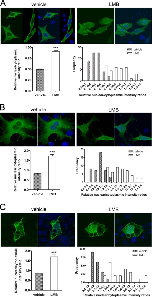
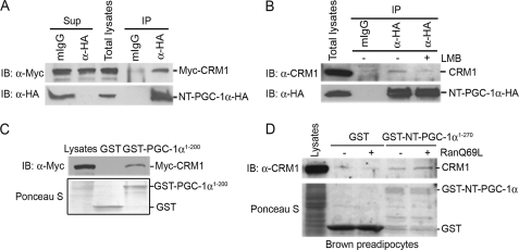
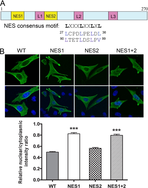
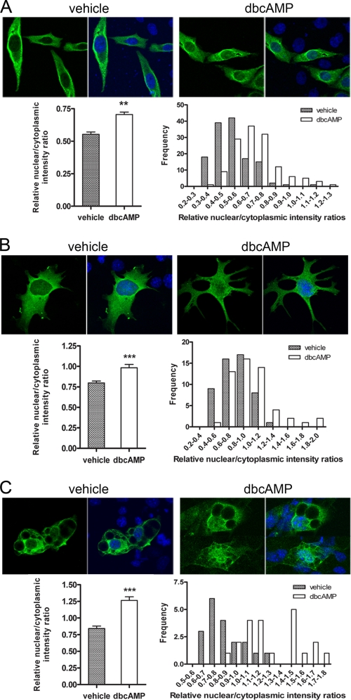
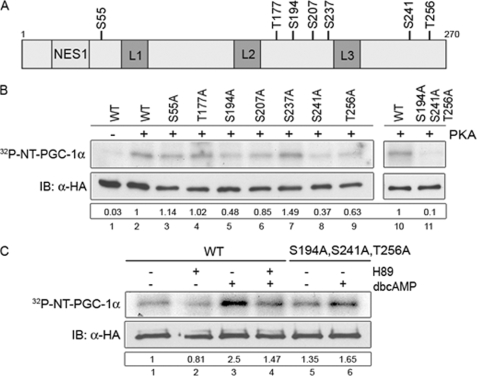
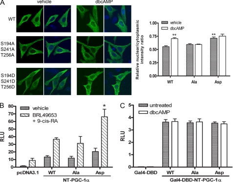
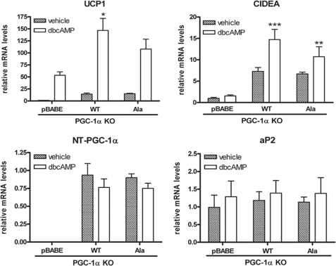
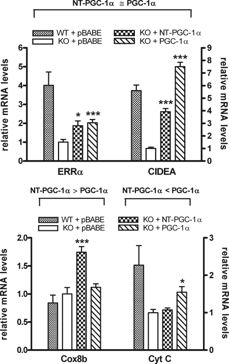
Peroxisome proliferator-activated receptor gamma co-activator-1alpha (PGC-1alpha) plays a central role in the regulation of cellular energy metabolism and metabolic adaptation to environmental and nutritional stimuli. We recently described a novel, biologically active splice variant of PGC-1alpha (NT-PGC-1alpha, amino acids 1-270) that retains the ability to interact with and transactivate nuclear hormone receptors through its N-terminal transactivation domain. Whereas PGC-1alpha is an unstable nuclear protein sensitive to ubiquitin-mediated targeting to the proteasome, NT-PGC-1alpha is relatively stable and predominantly cytoplasmic, suggesting that its ability to interact with and activate nuclear receptors and transcription factors is dependent upon regulated access to the nucleus. We provide evidence that NT-PGC-1alpha interacts with the nuclear exportin, CRM1, through a specific leucine-rich domain (nuclear export sequence) that regulates its export to the cytoplasm. The nuclear export of NT-PGC-1alpha is inhibited by protein kinase A-dependent phosphorylation of Ser-194, Ser-241, and Thr-256 on NT-PGC-1alpha, which effectively increases its nuclear concentration. Using site-directed mutagenesis to prevent or mimic phosphorylation at these sites, we show that the transcriptional activity of NT-PGC-1alpha is regulated in part through regulation of its subcellular localization. These findings suggest that the function of NT-PGC-1alpha as a transcriptional co-activator is regulated by protein kinase A-dependent inhibition of CRM1-mediated export from the nucleus.


Figures

FIGURE 1.
FIGURE 1.
Cytoplasmic localization of NT-PGC-1α is sensitive to LMB. A, translocation of NT-PGC-1α-HA from nucleus to the cytoplasm is inhibited following treatment with LMB in CHO-K1 cells. CHO-K1 cells were transiently transfected with NT-PGC-1α-HA and treated with vehicle alone or LMB for 1 h. Quantitative determination of the relative nuclear to cytoplasmic intensity ratios was performed as described under “Experimental Procedures.” The bar diagram of the relative ratios of nuclear to cytoplasmic fluorescence intensity of NT-PGC-1α is shown with the mean value and standard error. Significant deference by Student's t test is shown; ***, p < 0.0001 versus control. The nuclear to cytoplasmic ratio values were presented as frequency distribution. B, PGC-1α-null brown preadipocyte cells were adenovirally transduced with Ad-NT-PGC-1α-FLAG and treated with vehicle alone or LMB for 1 h. The bar diagram of the relative ratios of nuclear to cytoplasmic fluorescence intensity of NT-PGC-1α is shown with the mean ± S.D. Significant deference by Student's t test is shown; ***, p < 0.0001 versus control. The nuclear to cytoplasmic ratio values are presented as frequency distribution. C, fully differentiated PGC-1α-deficient brown adipocyte cells were transduced with Ad-NT-PGC-1α-FLAG and treated with vehicle alone or LMB for 1 h. The bar diagram of the relative ratios of nuclear to cytoplasmic fluorescence intensity of NT-PGC-1α is shown with the mean ± S.D. Significant deference by Student's t test is shown; ***, p < 0.0001 versus control. The nuclear to cytoplasmic ratio values are presented as frequency distribution.
FIGURE 2.
FIGURE 2.
Interaction of NT-PGC-1α with CRM1. A, coimmunoprecipitation of NT-PGC-1α-HA and Myc-CRM1 in transfected CHO-K1 cells. Immunoprecipitates with anti-HA antibody were separated by SDS-PAGE and immunoblotted with anti-Myc antibody (top). The same blot was reprobed with anti-HA antibody to confirm the presence of NT-PGC-1α-HA (bottom). B, coimmunoprecipitation of NT-PGC-1α-HA with endogenous CRM1 in CHO-K1 cells that express NT-PGC-1α-HA. Immunoprecipitates with anti-HA antibody were separated by SDS-PAGE and immunoblotted with anti-CRM1 antibody (top). The same blot was reprobed with anti-HA antibody to confirm the presence of NT-PGC-1α-HA (bottom). C, in vitro interaction of GST-PGC-1α (1–200) with Myc-CRM1. GST or GST-PGC-1α (aa 1–200) fusion proteins immobilized on the beads were incubated with the CHO-K1 cell lysates that express Myc-CRM1. GST pulldown assays were separated by SDS-PAGE and immunoblotted with anti-Myc antibody (top). Expression of GST and GST-PGC-1α (aa 1–200) is shown in Ponceau S staining (bottom). D, GST-NT-PGC-1α interacts with CRM1 in a RanGTP-regulated manner. GST or GST-NT-PGC-1α fusion proteins immobilized on the beads were incubated with brown preadipocyte cell lysates without or with addition of affinity-purified RanQ69L. GST pulldown assays were separated by SDS-PAGE and immunoblotted with anti-CRM1 antibody (top). Expression of GST and GST-NT-PGC-1α is shown in Ponceau S staining (bottom).
FIGURE 3.
FIGURE 3.
Identification of NES within NT-PGC-1α. A, schematic representation of the structure of NT-PGC-1α. Two putative NES consensus sequences (Lx(1–3)[livfm]x(2–3)lx[lIV]) are indicated. B, effects of single or double NES mutations (Leu/Val to Ala) on nuclear export of NT-PGC-1α, CHO-K1 cells were transiently transfected with NT-PGC-1α-HA, NT-PGC-1α-NES1-HA. NT-PGC-1α-NES2-HA, or NT-PGC-1α-NES1 plus 2-HA. The bar diagram of the relative ratios of nuclear to cytoplasmic fluorescence intensity is shown with the mean ± S.E. Significant deference by one-way analysis of variance is shown; ***, p < 0.0001 versus control.
FIGURE 4.
FIGURE 4.
Nuclear accumulation of NT-PGC-1α is increased by cAMP analogue. A, nuclear accumulation of NT-PGC-1α occurs following treatment with Bt2cAMP. CHO-K1 cells were transiently transfected with NT-PGC-1α-HA and treated with vehicle alone or Bt2cAMP for 1 h. The bar diagram of the relative ratios of nuclear to cytoplasmic fluorescence intensity of NT-PGC-1α is shown with the mean ± S.D. Significant deference by Student's t test is shown; **, p = 0.0044 versus control. The nuclear to cytoplasmic ratio values are presented as frequency distribution. B, nuclear accumulation of NT-PGC-1α following treatment with vehicle alone or Bt2cAMP in PGC-1α-null brown preadipocyte cells transduced with Ad-NT-PGC-1α-FLAG. The bar diagram of the relative ratios of nuclear to cytoplasmic fluorescence intensity of NT-PGC-1α is shown with the mean ± S.D. Significant deference by Student's t test is shown; **, p = 0.001 versus control. The nuclear to cytoplasmic ratio values are presented as frequency distribution. C, nuclear accumulation of NT-PGC-1α following treatment with vehicle alone or Bt2cAMP in PGC-1α-deficient fully differentiated brown adipocyte cells transduced with Ad-NT-PGC-1α-FLAG. The bar diagram of the relative ratios of nuclear to cytoplasmic fluorescence intensity of NT-PGC-1α is shown with the mean ± S.D. Significant deference by Student's t test is shown; ***, p < 0.0001 versus control. The nuclear to cytoplasmic ratio values were presented as frequency distribution.
FIGURE 5.
FIGURE 5.
PKA-dependent phosphorylation of NT-PGC-1α. A, schematic diagram shows seven putative PKA consensus phosphorylation sites ([R,K]XX[S,T]) in NT-PGC-1α. B, in vitro PKA-mediated phosphorylation of NT-PGC-1α. NT-PGC-1α-HA and its serine/threonine to alanine mutants were in vitro transcribed/translated using a TnT T7 Coupled Reticulocyte Lysate System, immunoprecipitated, and subjected for the in vitro kinase assay with the purified bovine catalytic subunit of PKA. Autoradiography (top) shows phosphorylated NT-PGC-1α by PKA. Western blot (bottom) shows expression of NT-PGC-1α-HA and its serine/threonine to alanine mutants. Numbers in the box represent the relative phosphorylation that is normalized by NT-PGC-1α protein. C, in situ PKA-dependent phosphorylation of NT-PGC-1α. Autoradiography (top) shows phosphorylated NT-PGC-1α in 32P-labeled CHO-K1 cells ectopically expressing NT-PGC-1α-HA (untreated and H89/Bt2cAMP-treated) and NT-PGC-1α-S194A/S241A/T256A-HA (untreated and Bt2cAMP-treated). Western blot (bottom) shows expression of NT-PGC-1α-HA and NT-PGC-1α-S194A/S241A/T256A-HA. Numbers in the box represent the relative phosphorylation that is normalized by NT-PGC-1α protein.
FIGURE 6.
FIGURE 6.
PKA-dependent regulation of subcellular localization and transcriptional activity of NT-PGC-1α. A, localization of NT-PGC-1α-HA, NT-PGC-1α-S194A/S241A/T256A-HA, and NT-PGC-1α-S194D/S241D/T256D-HA in CHO-K1 cells. The bar diagram of the relative ratios of nuclear to cytoplasmic fluorescence intensity is shown with the mean ± S.D. Significant deference by Student's t test; **, p < 0.01. B, transcriptional co-activation assay using a luciferase reporter assay. pcDNA3.1, NT-PGC-1α-HA, NT-PGC-1α-S194A/S241A/T256A-HA, or NT-PGC-1α-S194D/S241D/T256D-HA were co-transfected in CHO-K1 cells with a PPRE luciferase reporter (PPRE X3-TK-luc), RXRα, PPAPγ1, and a Renilla luciferase reporter. 24 h after transfection cells were treated with vehicle or with BRL49653 and 9-cis-retinoic acid. Luciferase activity was determined 48 h after transfection and the relative luciferase units were calculated as described under “Experimental Procedures.” Data represent mean ± S.D. of at least three independent experiments. Significant deference by Student's t test: *, p < 0.05. C, transcriptional activation assay. Gal4-DBD, Gal4-NT-PGC-1α, Gal4-NT-PGC-1α-S194A/S241A/T256A, and Gal4-NT-PGC-1α-S194D/S241D/T256D were co-transfected in CHO-K1 cells with a luciferase reporter containing Gal4 DNA binding sites and a Renilla luciferase reporter. Luciferase activity was determined as above. Data represent mean ± S.D. of at least five independent experiments.
FIGURE 7.
FIGURE 7.
NT-PGC-1α-dependent induction of UCP1 and CIDEA is reduced by mutation of sites phosphorylated by PKA. UCP1, CIDEA, aP2, and NT-PGC-1α mRNA levels were assessed by real-time PCR analysis in differentiated PGC-1α KO brown adipocyte cells where pBABE, NT-PGC-1α-HA, or NT-PGC-1α-S194A/S241A/T256A-HA was retrovirally expressed. On the seventh day of differentiation, cells were treated with or without Bt2cAMP for 4 h. Relative abundance of mRNA was normalized to cyclophilin mRNA. Data represent mean ± S.D. of at least three independent experiments. Significant difference of UCP1 and CIDEA gene expression was determined by Student's t test; *, p < 0.05; **, p < 0.01; and ***, p < 0.001.
FIGURE 8.
FIGURE 8.
Genes that are comparably (top panel) or differentially (bottom panel) regulated by NT-PGC-1α and PGC-1α. ERRα, CIDEA, Cox8b, and CytC mRNA levels were assessed by real-time PCR in differentiated brown adipocytes from either wild-type or PGC-1α null mice. On day 7 of differentiation, wild-type cells expressing empty vector (pBABE) or PGC-1α null cells expressing empty vector, NT-PGC-1α, or PGC-1α were treated with Bt2cAMP for 4 h. Relative abundance of mRNA was normalized to cyclophilin mRNA. Data represent mean ± S.D. of at least three independent experiments. Significant deference by Student's t test is shown; *, p < 0.05 and ***, p < 0.001.

References

    1. Finck B. N., Kelly D. P. (2006) J. Clin. Invest. 116, 615–622 - PMC - PubMed
    1. Handschin C., Spiegelman B. M. (2006) Endocr. Rev. 27, 728–735 - PubMed
    1. Puigserver P., Wu Z., Park C. W., Graves R., Wright M., Spiegelman B. M. (1998) Cell 92, 829–839 - PubMed
    1. Wu Z., Puigserver P., Andersson U., Zhang C., Adelmant G., Mootha V., Troy A., Cinti S., Lowell B., Scarpulla R. C., Spiegelman B. M. (1999) Cell 98, 115–124 - PubMed
    1. Lehman J. J., Barger P. M., Kovacs A., Saffitz J. E., Medeiros D. M., Kelly D. P. (2000) J. Clin. Invest. 106, 847–856 - PMC - PubMed

Publication types

MeSH terms

Substances

LinkOut - more resources