. 2010 May;24(5):993-1006.
doi: 10.1210/me.2009-0438. Epub 2010 Mar 29.

Xenoestrogen-induced regulation of EZH2 and histone methylation via estrogen receptor signaling to PI3K/AKT

Affiliations

Xenoestrogen-induced regulation of EZH2 and histone methylation via estrogen receptor signaling to PI3K/AKT

Tiffany G Bredfeldt et al. Mol Endocrinol. 2010 May.

Abstract

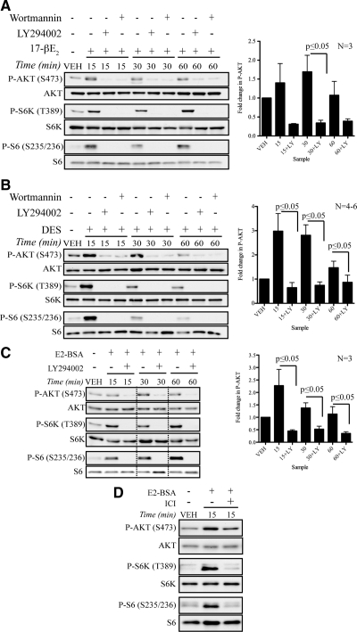
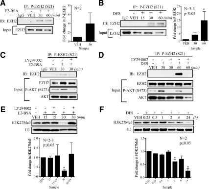
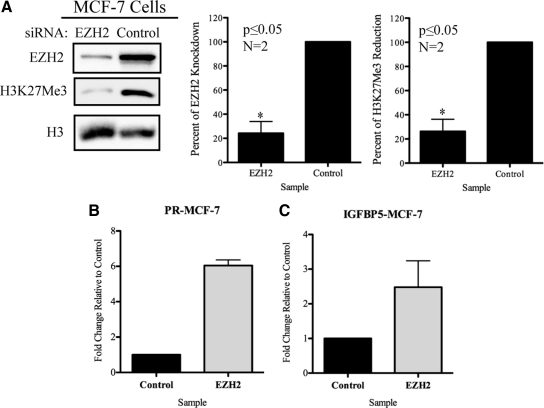
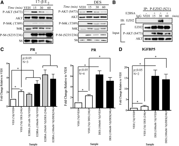
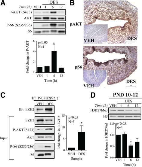
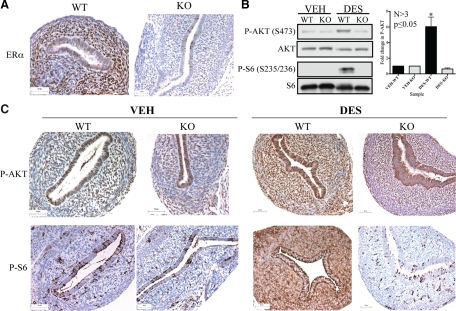
Although rapid, membrane-activated estrogen receptor (ER) signaling is no longer controversial, the biological function of this nongenomic signaling is not fully characterized. We found that rapid signaling from membrane-associated ER regulates the histone methyltransferase enhancer of Zeste homolog 2 (EZH2). In response to both 17beta-estradiol (E2) and the xenoestrogen diethylstilbestrol, ER signaling via phosphatidylinositol 3-kinase/protein kinase B phosphorylates EZH2 at S21, reducing levels of trimethylation of lysine 27 on histone H3 in hormone-responsive cells. During windows of uterine development that are susceptible to developmental reprogramming, activation of this ER signaling pathway by diethylstilbestrol resulted in phosphorylation of EZH2 and reduced levels of trimethylation of lysine 27 on histone H3 in chromatin of the developing uterus. Furthermore, activation of nongenomic signaling reprogrammed the expression profile of estrogen-responsive genes in uterine myometrial cells, suggesting this as a potential mechanism for developmental reprogramming caused by early-life exposure to xenoestrogens. These data demonstrate that rapid ER signaling provides a direct linkage between xenoestrogen-induced nuclear hormone receptor signaling and modulation of the epigenetic machinery during tissue development.


Figures

Figure 1
Figure 1
ER-mediated activation of PI3K/AKT signaling. A, AKT activation by E2 (50 nm) can be inhibited by pretreatment with PI3K antagonists, LY294002 (20 μm) and wortmannin (200 nm), in MCF-7 cells. B, DES-induced activation of AKT pathway is inhibited by PI3K antagonists, LY294002 (20 μm) and wortmannin (200 nm), which were given to some groups of MCF-7 cells before DES (50 nm) treatment. C, MCF-7 cells were pretreated with LY294002 (20 μm) and exposed to E2-BSA (25 nm) for described times. Figure was cropped as indicated by dotted lines. D, E2-BSA (25 nm)-induced nongenomic signaling is inhibited by pretreatment with ICI 182780 (100 nm) in MCF-7 cells. Each panel shows representative Western blots from an independent experiment. Graphs display densitometry analysis of the ratio of phospho-AKT to total AKT protein normalized to vehicle-treated cells (mean ± sem). Statistical significance (*, P ≤ 0.05) was determined by Student’s t test. LY, LY294002.
Figure 2
Figure 2
Nongenomic signaling to PI3K/AKT phosphorylates EZH2 and reduces H3K27Me3 levels. A, Nongenomic ER signaling to the AKT pathway increases P-EZH2 levels in E2-BSA (25 nm)-treated lysates as detected by immunoprecipitation in MCF-7 cells. Western blots of inputs from immunoprecipitation were used to control for variation in total EZH2 in each sample. B, DES (50 or 100 nm) induces phosphorylation of endogenous EZH2 in MCF-7 cells. C, Immunoprecipitation of P-EZH2 from MCF-7 cell-treated E2-BSA (25 nm) with or without LY294002 (20 μm) pretreatment demonstrates dependence of EZH2 phosphorylation on PI3K/AKT pathway. D, DES-induced (50 or 100 nm) phosphorylation of endogenous EZH2 is dependent on the activity of the PI3K/AKT pathway. E, Western blot analysis of MCF-7 cells treated with E2-BSA (25 nm) with or without LY294002 (20 μm) demonstrates dependence of the reduction of H3K27Me3 levels on PI3K-mediated AKT activation. F, Histones were acid precipitated from estrogen-deprived MCF-7 cells that were treated with VEH or DES (50 nm) for 0.25, 0.5, 1, 2, 6, and 24 h and subjected to Western blot analysis, revealing that DES reduces H3K27Me3 levels in chromatin of treated cells. Western blots from independent experiments were analyzed by densitometry, and differences in levels of phosphorylation or methylation relative to VEH are shown as mean ± sem. Statistical significance was determined by Student’s t test or one-way ANOVA (*, P ≤ 0.05). IB, Immunoblotting; IP, immunoprecipitation; LY, LY294002.
Figure 3
Figure 3
EZH2 is the primary HMT responsible for H3K27Me3 and is a regulator of PR and IGFBP5 genes. A, Knockdown of EZH2 protein reduces levels of H3K27Me3 in MCF-7 cells. B, EZH2 is a regulator of PR in MCF-7 cells. RNA was collected from cells treated with EZH2 or control siRNA, and PR expression levels were analyzed by qPCR. C, IGFBP5 is an EZH2 target gene as detected by qPCR analysis of MCF-7 cells where EZH2 protein levels are reduced by siRNA. Panels display a representative Western blot from two independent experiments. Western blot data investigating efficiency of EZH2 knockdown and subsequent reduction in H3K27Me3 levels were analyzed by densitometry. Significance was given to data with P ≤ 0.05 (*) as identified by Student’s t test. For qPCR analysis the calibrator of this experiment was cells treated with control siRNA. Fold change was calculated for each sample in comparison with the calibrator. Graph bars represent mean ± sem obtained from a single experiment read in triplicate.
Figure 4
Figure 4
Estrogens activate nongenomic signaling to phosphorylate EZH2 and regulate gene expression in ELT3 cells. A, ELT3 cells were starved for 48 h and then treated with E2 (10 nm) or DES (10 nm) for indicated times. Activation of PI3K/AKT pathway was determined by Western blot analysis. B, Activation of PI3K/AKT pathway by E2-BSA (50 or 100 nm) induces EZH2 phosphorylation in ELT3 cells, which was detected by immunoprecipitation followed by Western blot analysis. C, ELT3 cells exposed to VEH, E2-BSA (50 or 100 nm), or DES (100 nm) for 7 d. After removal of initial, priming treatments cells were washed with PBS and starved (48 h), cells were reexposed to DES (50 nm), and expression of PR was analyzed by qPCR demonstrating that E2-BSA and DES were capable of reprogramming the basal expression and hormone responsiveness of this gene. D, ELT3 cells were exposed to VEH or DES (100 nm) for 7 d. Treatment media were removed, and cells were washed with PBS and replaced with serum- and estrogen-free medium. After 48 additional hours, cells were rechallenged with DES (50 nm) or VEH (ethanol) for 24 h. Expression of the IGFBP5 gene was analyzed by qPCR, revealing that DES reprogrammed the basal expression level and hormone responsiveness of this gene relative to VEH-primed cells. Bar graphs display mean ± sem gene expression relative to VEH-treated cells from two to four independent experiments. Student’s t test was used to determine statistical significance (*, P ≤ 0.05). IB, Immunoblotting; IP, immunoprecipitation.
Figure 5
Figure 5
Xenoestrogen-induced modulation of H3K27Me3 in vivo. A, Uteri were pooled from two to three Eker rats injected with VEH or DES. Western blot analysis of PI3K/AKT pathway demonstrated activation of nongenomic signaling in vivo by DES. B, Uteri were collected from Eker rats exposed to DES or VEH for 6 h and stained for P-AKT and P-S6, revealing activation of PI3K/AKT in DES-treated tissue (n = 43 animals; 24 VEH and 19 DES). Scale bar, 75 μm. C, Whole uteri were pooled from one to three Eker rats and homogenized after 6 h DES or VEH exposure. Lysates were immunoprecipitated with anti-P-EZH2 antibody. Immunoprecipitates were analyzed by Western blot for total EZH2, demonstrating enrichment of P-EZH2 in DES-treated tissue. Inputs show activation of nongenomic signaling in treated animals. D, Uteri were pooled from two to three DES- or VEH-treated rats. Histones were acid precipitated. Western blot analysis of histone proteins reveals decreased H3K27Me3 levels after DES (1, 6, and 12 h) exposure relative to VEH-treated animals in animals exposed on PND 10–12, a window of development susceptible to developmental reprogramming. Densitometry analyses of Western blot data show differences in levels of phosphorylation or methylation relative to VEH. The bars in each graph represent mean ± sem. Statistical significance was determined by Student’s t test (*, P ≤ 0.05). IB, Immunoblotting; IP, immunoprecipitation.
Figure 6
Figure 6
Activation of PI3K/AKT signaling in the uterus is ERα mediated. A, Histological analysis of sections collected from uteri of ERKO or WT mice demonstrate that ERKO animals do not express ERα. Scale bar, 50 μm. B, Western blot analysis of uterine lysates from ERKO mice vs. WT mice treated as described above demonstrates that activation of the PI3K/AKT pathway is ERα dependent in vivo. Each lane represents lysates from one to two pooled uteri from animals of matched genotype. C, Histological analysis of ERKO or WT uteri exposed to VEH or DES (6 h) demonstrates that activation of nongenomic signaling is dependent on ERα. Scale bar, 50 μm. The panels in this figure display representative Western blots. Densitometry analysis of Western blot data presented in bar graph represents mean ± sem. Statistical significance was determined by Student’s t test (*, P ≤ 0.05).

Similar articles

Cited by

References

    1. Acconcia F, Kumar R 2006 Signaling regulation of genomic and nongenomic functions of estrogen receptors. Cancer Lett 238:1–14 - PubMed
    1. Gruber CJ, Tschugguel W, Schneeberger C, Huber JC 2002 Production and actions of estrogens. N Engl J Med 346:340–352 - PubMed
    1. Deroo BJ, Korach KS 2006 Estrogen receptors and human disease. J Clin Invest 116:561–570 - PMC - PubMed
    1. Nagel SC, vom Saal FS, Welshons WV 1999 Developmental effects of estrogenic chemicals are predicted by an in vitro assay incorporating modification of cell uptake by serum. J Steroid Biochem Mol Biol 69:343–357 - PubMed
    1. Colborn T, vom Saal FS, Soto AM 1993 Developmental effects of endocrine-disrupting chemicals in wildlife and humans. Environ Health Perspect 101:378–384 - PMC - PubMed

Publication types

MeSH terms